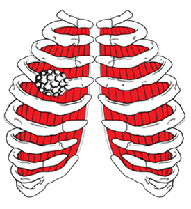
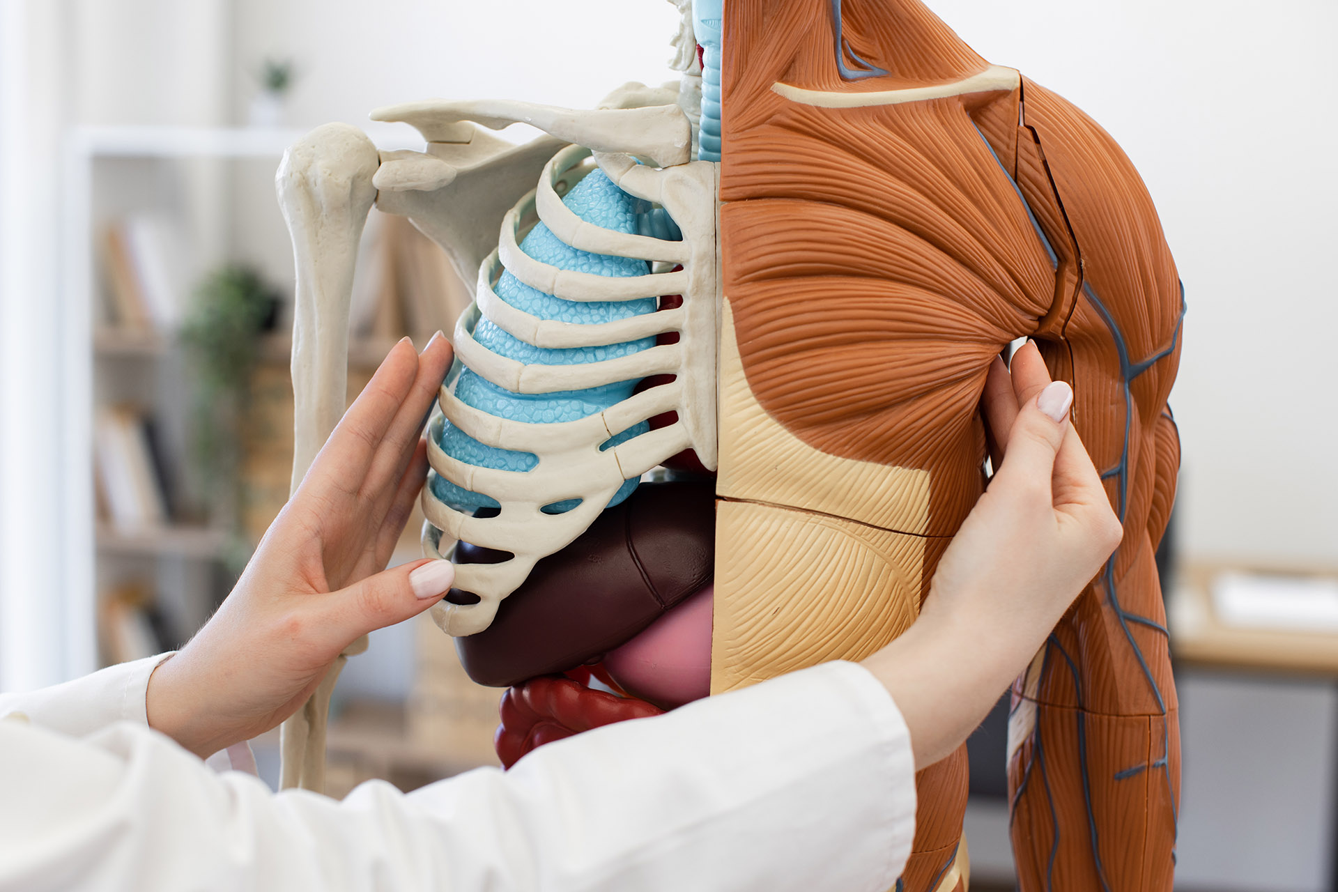
The chest wall creates a protective ‘cage’ to keep internal organs, such as the heart and lungs, safe. However, as in other parts of the body, your bones and tissues can also develop tumours on your chest wall. The chest cavity, also known as the thoracic cavity, is the anatomical space protected by the chest wall, housing vital organs such as the heart, lungs, and oesophagus. The chest wall forms the boundaries of this space, and its anatomy comprises bones, muscles, and blood vessels that play a crucial role in protecting the chest cavity.
The pathology of a chest wall tumour often depends on its origin and involvement in the chest wall structures. Tumours in the chest wall may arise from various tissues, and the space within the thoracic region is significant for containing these vital organs and potential tumour development.
Many different types of tumours can grow in this area, either stemming from the chest wall itself or spreading from other parts of the body. Bone tumours, such as osteosarcomas and chondrosarcomas, are among the types that can originate in the bone of the chest wall. A tumour that stems from the ribs or sternum is considered a primary chest wall tumour – a malignant neoplasm – and may or may not be cancerous. Common non-cancerous tumours include chondromas, osteochondromas and fibrous dysplasia of the rib. Tumours in the chest wall can also involve soft tissue or cartilage.
Metastasis may occur when cancer cells from the primary chest wall tumour spread to other areas of the body. These secondary neoplasms share the same type of cancer as the primary malignancy. Tumours can invade blood vessels, allowing cancer cells to enter the circulation and spread to distant sites.
The most common cancerous tumours that arise from the chest wall are the sarcomas. A sarcoma is the most common malignant tumour of the chest wall, forming in the bones, soft tissue or cartilage. A large number of malignant wall chest tumours are sarcomas, though their symptoms vary depending on the tumour’s classification and its severity. Some people can experience anything from difficulty breathing and pain to swelling surrounding the sarcoma.
Symptoms of chest tumours can be variable. Often, they are subtle at first and may go unnoticed. A tumour may present with a growing mass or with persistent pain in the chest wall, depending on its size, location and involvement of surrounding structures.
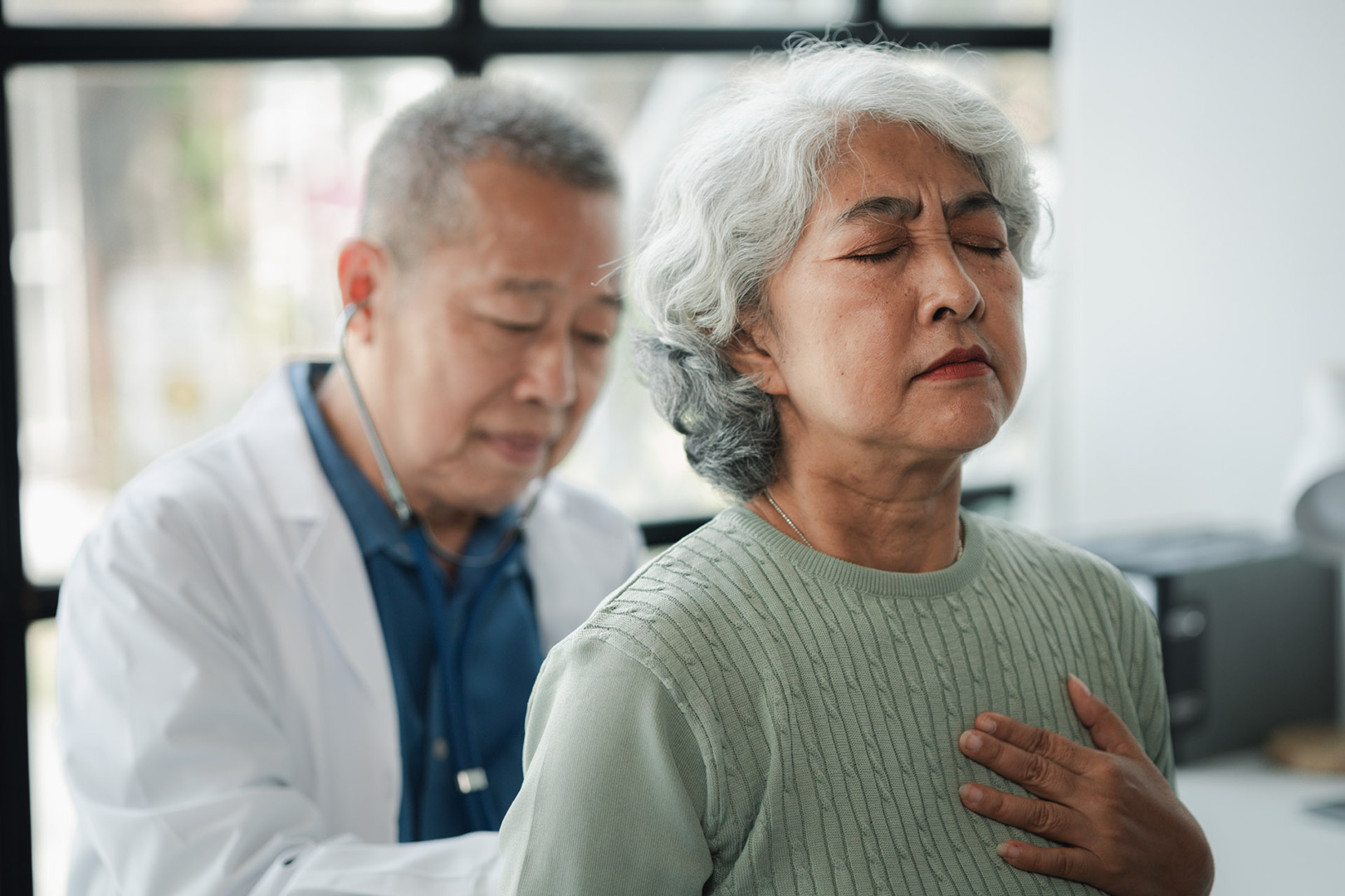
Symptoms of chest wall tumours often include swelling, pain and changes in chest shape.
Chest wall tumours have a variety of different symptoms, depending on their severity and classification. Sometimes, they present as mild lumps that are firm to the touch; other times, as masses that cause discomfort, pain and difficulty breathing. Occasionally, chest wall tumours can change the shape of the chest wall and may only be visible through imaging protocols. They can also present as a growing mass or cause pain, and may require a multidisciplinary approach for management.
Chest tumour symptoms may include fevers, night sweats, and/or weight loss, as it can affect other parts of the body.
Different types of chest wall tumours occur more frequently in men than in women. Chest wall tumours include both primary and secondary tumours, with diverse origins such as benign or malignant growths, infectious or inflammatory processes, and metastases from other organs. Some of these types tend to be more aggressive, while the types of tumours in the chest of females are more often benign. Ewing sarcoma is a common malignant tumour of the chest wall in children and young adults. Either way, there is a risk of a tumour being malignant, so it is important to get checked if you notice a new bump in your chest area.
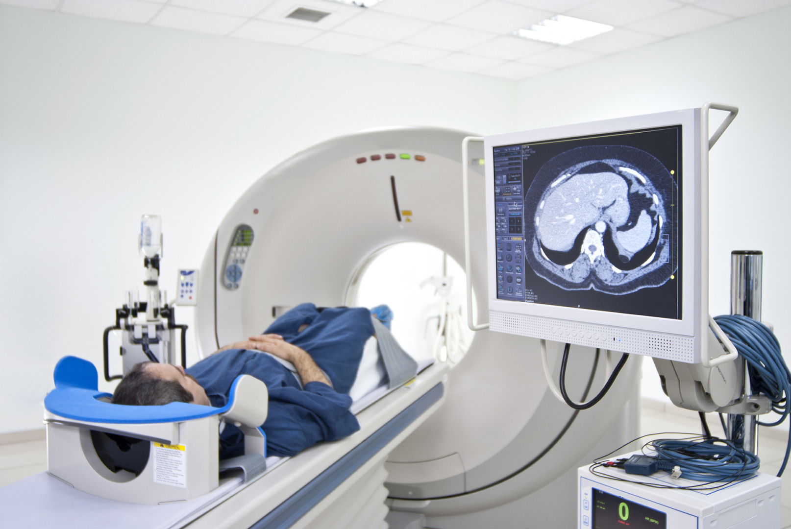
To check for abnormalities, our team may conduct chest X-rays, which are often the first imaging study performed for chest wall tumours. If an abnormality is found, additional tests such as a computed tomography (CT) scan or magnetic resonance imaging (MRI) will be performed to ascertain the tumour’s classification and severity. After these preliminary assessments are completed, a biopsy may be performed to diagnose the tumour and determine whether it is benign or malignant.
Chest wall tumours and sarcomas comprise a broad group of growths that can be either benign or malignant, with sarcomas representing one of the most common malignant types in this category. Sarcomas arise in the soft tissues, bones, or cartilage of the chest wall and are therefore considered a subset of malignant chest wall tumours.
Both chest wall tumours and sarcomas can present with similar symptoms, including pain, swelling or difficulty breathing if the tumour presses on nearby organs. Diagnosis often requires a combination of imaging and biopsy, not only to identify the tumour’s size and location but also to determine whether it is benign or malignant.
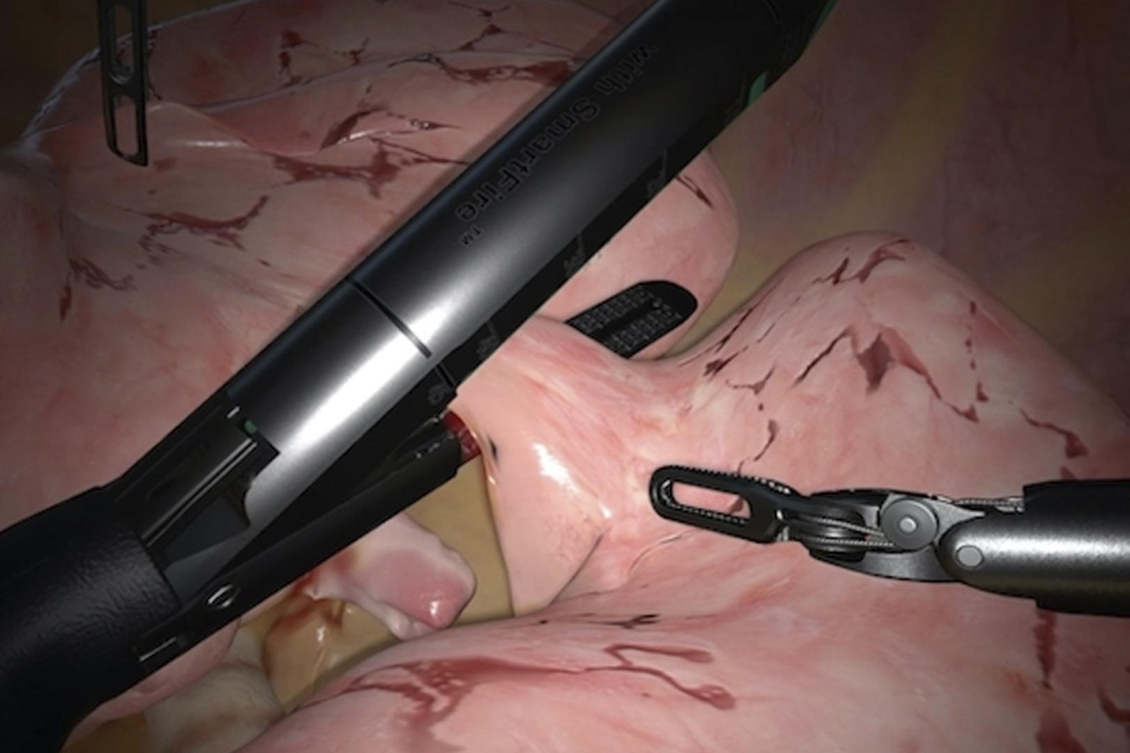
The overlap between these conditions is important. While not every chest wall tumour is a sarcoma, many sarcomas present as chest wall tumours. Treatment usually involves chest wall resection, which is a key component of surgical management for malignant tumours, ensuring complete oncologic excision with appropriate margins. For malignant sarcomas, additional therapies such as chemotherapy or radiotherapy may be recommended.
Thoracic surgery plays a crucial role in the multidisciplinary approach to the resection and reconstruction of chest wall tumours, helping to manage defect repair and preserve function after tumour removal.
Treatment of tumours depends on the type found, and benign chest wall tumours may be kept under observation. However, some benign chest wall masses may still need surgical resection.
Some rare chest wall tumours, such as solitary plasmacytoma, are composed of monoclonal plasma cells and may be treated with radiation therapy instead of surgery.
If the tumour is cancerous, treatment options may involve surgery, chemotherapy and possible radiotherapy. Surgical resection will include removing the entire tumour and reconstructing the chest wall with the adjacent muscles and titanium rib plates.
Recovery from chest wall tumour surgery varies, as many variables contribute to the body’s healing. The general rule is to expect a short hospital stay for observation, typically lasting a few days, following chest wall tumour surgery. After a few weeks, patients often resume their normal daily activities at the discretion of their healthcare provider. It usually takes several months for a full recovery.