

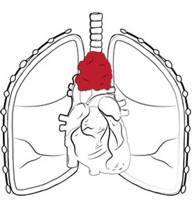
Different tumours arise from different tissues, some from fibrous tissue, connective tissues, or germ cell tissue, and some from immune system cells or lymph nodes. Some of these growths are benign tumours, such as thymic cysts, pericardial cysts, or slow-growing neurogenic tumours. Others are malignant tumours, including thymoma and thymic carcinoma, Hodgkin lymphoma and other lymphomas, and mediastinal germ cell tumours.
Mediastinal tumours are rare, occurring in less than 1% of the population, and most are found in adults aged 30 to 50. In children, masses are more often found in the posterior mediastinum and are frequently neurogenic tumours. In Singapore, the estimated incidence is 2.4 cases per 100,000 people, reflecting the local rarity of the disease.
“What causes a mediastinal mass?”, you may ask. The answer depends on the underlying pathology. Some masses arise from uncontrolled tumour cells, some from congenitally developed atypical tissue, and some from inflammation, infection, or enlarged lymph nodes. Risk factors vary by tumour type. Autoimmune diseases such as myasthenia gravis and rheumatoid arthritis are associated with thymoma and thymic carcinoma. Hodgkin lymphoma risk factors include prior Epstein-Barr virus infection and immune system dysfunction. That is why the phrase ‘mediastinal tumour’ describes a location rather than a single cause.
Mediastinal mass symptoms often result from pressure on nearby structures, but many patients have no symptoms at all. When a tumour grows large enough, symptoms often reflect compression of the airway, oesophagus, nerves, heart, or large blood vessels. Up to about 40% with mediastinal tumours do not notice symptoms and are diagnosed incidentally on a chest X-ray or CT scan. Malignant tumours are more likely to be symptomatic than benign tumours.
Typical mediastinal tumour symptoms include a cough, shortness of breath, wheezing, chest pain or fullness, difficulty swallowing, hoarseness, fever, chills, night sweats, coughing up blood, tender or swollen lymph nodes, and unintentional or an unexplained weight loss.
Symptoms can also vary by the compartment and tumour type. Anterior mediastinal tumours and an anterior mediastinal mass may cause a cough, shortness of breath, chest discomfort, and obstruction of the superior vena cava, with facial swelling and distended neck veins. Hodgkin lymphoma presenting as an anterior mediastinal mass can also cause a fever, night sweats, and unexplained weight loss.
Posterior mediastinal tumours are more likely to cause back pain, spinal cord compression, neurological symptoms, or swallowing problems because they lie close to the spinal column, nerve roots and the oesophagus. Some thymoma and thymic carcinoma tumours may be linked with autoimmune diseases or paraneoplastic syndromes such as myasthenia gravis.
You should see a lung specialist or thoracic surgeon promptly if imaging done shows a mediastinal mass, or if you have persistent chest symptoms that do not settle.
Any unexplained chest pain, persistent cough, shortness of breath, hoarseness, difficulty swallowing, or facial swelling warrants a thorough review, especially if imaging has already revealed a mass. Even a benign tumour can become serious because of where it sits. Untreated mediastinal tumours can press on the heart, pericardium, spinal cord, trachea, or surrounding vascular structures, causing serious complications. This is why a mediastinal mass can sometimes be life-threatening even when it is not cancer.
Seeing a thoracic surgeon early does not mean surgery will definitely be needed. It means the mass should be assessed by someone who understands the anatomy, the likely differential diagnosis, and the safest way to obtain adequate tissue if a biopsy is needed. At Neumark Lung & Chest Surgery Centre, this includes deciding whether the next step is surveillance, a biopsy, chemotherapy and radiation, targeted therapy, or mediastinal tumour surgery using a minimally invasive approach where appropriate.
Non-surgical treatment of mediastinal masses depends on the tumour type. In some cases, a non-surgical approach is the primary treatment rather than a fallback.
In other words, any treatment for a mediastinal mass is driven by tissue diagnosis, anatomy, and resectability, not by the tumour size alone.
An inoperable mediastinal tumour is one that cannot be removed safely or completely because of the tumour’s extent, invasion of vital structures, or the nature of the disease. In that setting, treatment may focus on chemotherapy and radiation, targeted therapy, symptom relief, or combined oncological care with radiation oncologists. These can shift the focus from immediate resection to disease control and quality of life.
People often look for a single number for mediastinal tumour survival rate, but there is no single figure that applies to all mediastinal masses. The same goes for mediastinal tumour life expectancy, as a benign bronchogenic cyst has a very different outlook from Hodgkin lymphoma, thymic carcinoma, or a malignant neurogenic tumour. There is no single mediastinal tumour growth rate either. Hodgkin lymphoma can enlarge much faster, while a thymoma is often slower-growing, which is one reason the exact pathology matters so much. Even within one category, the stage, completeness of resection, tumour biology, and response to treatment matter greatly.
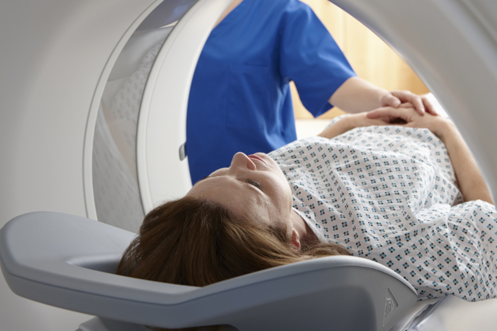
Mediastinal mass surgery is often the treatment of choice for resectable tumours, symptomatic mediastinal cysts and selected malignant tumours.
The operation depends on the tumour compartment, suspected pathology, tumour size, and the tumour’s relationship to surrounding structures and tissue. Surgery may be performed for a variety of reasons, such as to remove benign tumours or a thymic cyst, resect a thymoma and thymic carcinoma, clear a residual germ cell mass after chemotherapy, or obtain a definitive diagnosis when a less invasive biopsy is not enough. The surgical aim is a complete and safe resection while preserving vital structures wherever possible. Here are several modern surgical techniques designed to treat mediastinal masses as non-invasively as possible.
Robotic-Assisted Thoracic Surgery (RATS) is especially useful in the narrow, crowded space of the mediastinum. RATS offers 3D visualisation, fine instrument control, and precise dissection through a few small incisions. These features can be particularly helpful when tumours develop near the heart, trachea, oesophagus, and thoracic inlet, and may reduce blood loss, pain, and hospital stay for selected patients.
Video-Assisted Thoracoscopic Surgery (VATS) remains a very important minimally invasive option for well-defined, smaller lesions, especially in the middle and posterior mediastinum. It allows the surgeon to work through small keyhole incisions using a thoracoscopic camera and long instruments, avoiding a full thoracotomy in many patients. For select benign neurogenic tumours, thymic cysts, pericardial cysts and mediastinal cysts, VATS can offer less invasive trauma, quicker mobilisation, and fewer wound complications.
Uniportal VATS (U-VATS) uses a single incision and is part of Neumark’s broader minimally invasive thoracic practice for lung, mediastinal and chest wall conditions. It may be suitable for selected mediastinal lesions in which single-port access can provide safe exposure and effective dissection. This choice depends on tumour position, complexity, and the surgeon’s experience.
Open surgery still has an important role for very large tumours, masses invading the thoracic inlet or connective tissues, or cases requiring reconstruction.
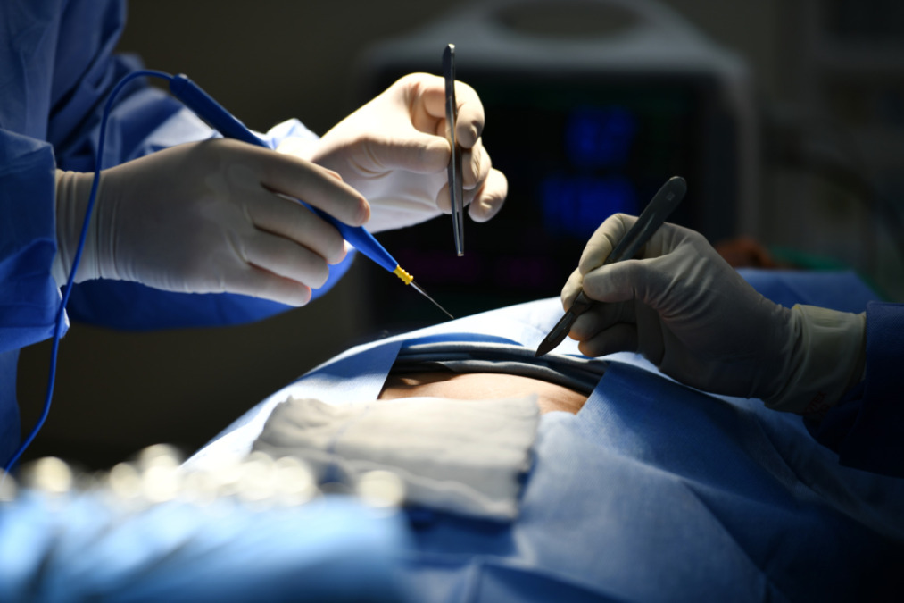
Recovery after mediastinal tumour treatment depends on the pathology, the extent of surgery, and whether chemotherapy and radiation or targeted therapy are also needed.
After minimally invasive surgery, many patients are up and moving early and leave the hospital sooner than after open surgery. Even so, surgery still carries risks, such as bleeding, air leak, infection, injury to nearby nerves or vessels, chest pain, incomplete resection, and the need to convert to an open operation if safety requires it.
Follow-up is essential because recurrence of a mediastinal mass depends on the final diagnosis and the extent of lesion removal. Some benign tumours, such as thymic and pericardial cysts, can be cured with surgery. Malignant or incompletely resected tumours may need surveillance with periodic CT scans or PET, and some patients need adjuvant chemotherapy and radiation or targeted therapy to reduce recurrence risk. Hodgkin lymphoma recurrence is monitored with PET and bone marrow assessment as needed.
Neumark Lung & Chest Surgery Centre specialises in minimally invasive thoracic surgery with a multidisciplinary approach led by Dr Harish Mithiran, senior consultant thoracic surgeon at Gleneagles and Mount Alvernia hospitals.
For patients with mediastinal tumours and mediastinal masses, this means a careful review of chest X-rays, CT scans, and MRI findings, thoughtful selection of biopsy techniques to obtain adequate tissue, and tailored planning for observation, medical therapy, and/or surgery. The centre’s thoracic practice includes experience with RATS, VATS, and U-VATS across a range of chest and mediastinal conditions.
If you have been told you have a mediastinal mass, or if you have ongoing chest pain or shortness of breath without a clear explanation, contact Neumark for a consultation. Early specialist review can clarify the diagnosis, identify the safest next step, and help you move forward with a plan that protects both your health and your quality of life.