

What Are Desmoid Tumours?
Desmoid tumours, also known as aggressive fibromatosis or desmoid fibromatosis, are rare, noncancerous growths that originate from connective tissues. These tumours belong to a group of rare, locally aggressive soft-tissue tumours known as fibromatoses, characterised by their potential to invade adjacent structures but without metastasis.
When occurring on the chest wall, which accounts for approximately 10% of cases, desmoid tumours pose unique challenges due to their proximity to vital organs, such as the lungs, heart and major blood vessels. Predominantly affecting young adults in their 20s and 30s, these tumours are more common in women, suggesting a possible hormonal influence. Early diagnosis is critical to tailor treatments and minimise complications in chest wall desmoid tumours.
Cellular and Risk Factors
Desmoid chest wall tumours often arise from mutations in the β-catenin gene, which drives the overproliferation of spindle-shaped cells, forming dense, fibrous masses. Risk factors include genetic predispositions, such as familial adenomatous polyposis (FAP) or Gardner syndrome, which significantly increase the likelihood of tumour development. Hormonal factors, like elevated estrogen levels during pregnancy or oral contraceptive use, and prior trauma or surgery in the chest area, can also trigger desmoid tumour formation.
Types of Desmoid Tumours in the Chest Wall
Desmoid tumours are exceedingly rare, with an annual incidence of 2–4 cases per million people. Of that small number, only 10–20% of these tumours occur in the chest wall, making them a unique clinical challenge. Their rarity underscores the need for specialised thoracic expertise.
Desmoid tumours in the chest wall can present in several distinct locations, each with unique characteristics and implications for treatment. The most common locations include extra-abdominal desmoid tumours, which develop outside the abdominal cavity and frequently involve the chest wall, leading to symptoms such as chest pain, swelling and restricted movement.
Chest wall fibromatosis is a specific type that targets the tissues of the chest wall, often resulting in persistent discomfort and, in some cases, breathing difficulties due to the tumour’s proximity to vital structures.
Desmoid-type fibromatosis is particularly notable for its aggressive local behaviour and high recurrence rates, making ongoing monitoring essential. Spindle cell desmoid tumours, composed primarily of spindle-shaped cells, can also arise in the chest wall and are associated with pain and swelling.

Recognising Symptoms
Chest wall desmoid tumours often present with subtle but persistent symptoms. Patients may experience dull, aching chest pain that worsens with movement or deep breathing, alongside an unnerving, firm, immovable mass under the skin. Swelling is common, and in advanced cases, tumours may compress airways, causing respiratory distress. For example, a typical patient may present with a slowly enlarging chest wall mass, localised pain and restricted arm movement. An imaging test may reveal tumour invasion into adjacent soft tissue.
Beyond physical symptoms, these tumours can lead to anxiety, depression and functional limitations, such as difficulty with arm movement or daily tasks. Early recognition of these signs is essential for proactive management and preserving quality of life.

Diagnostic Approaches
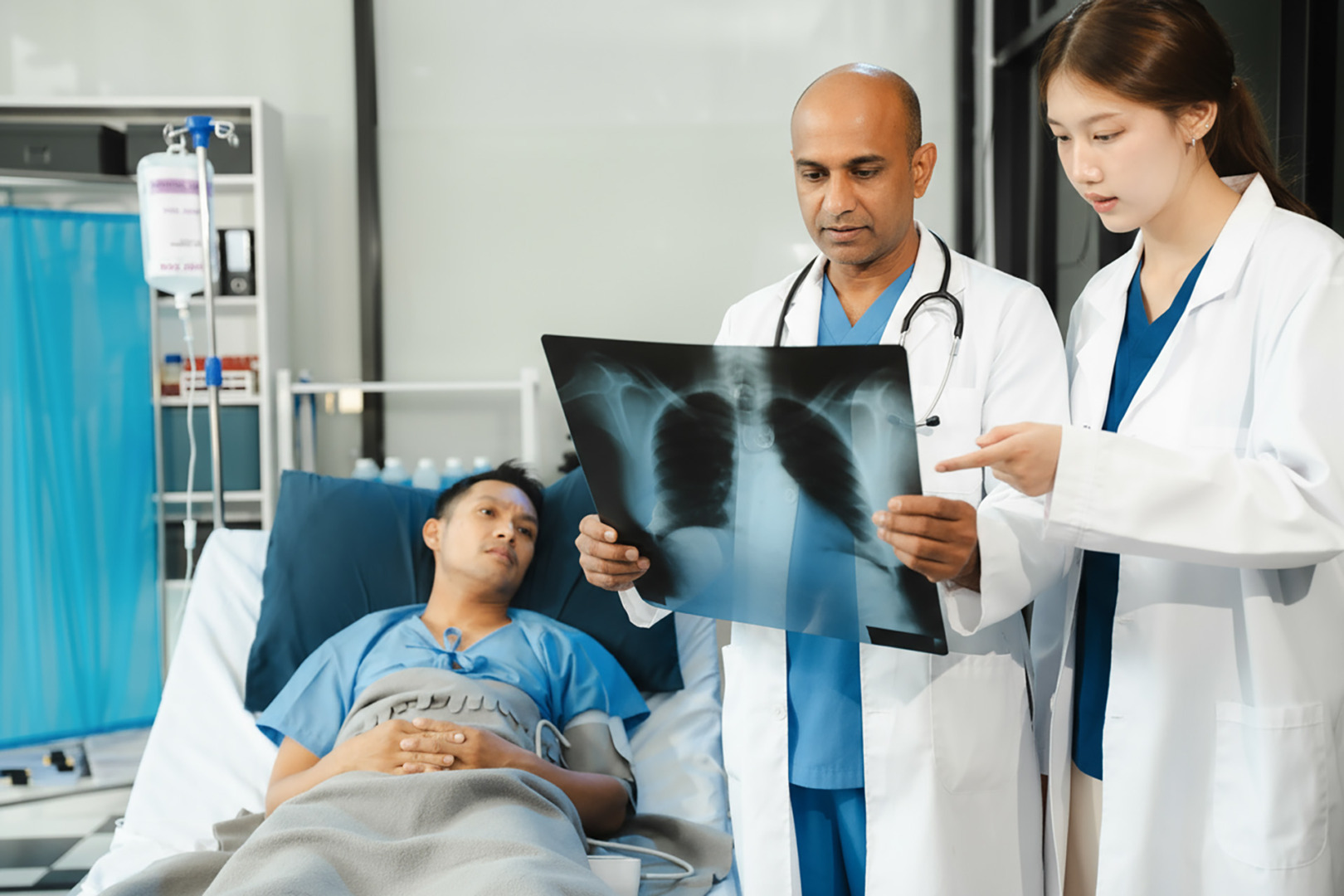
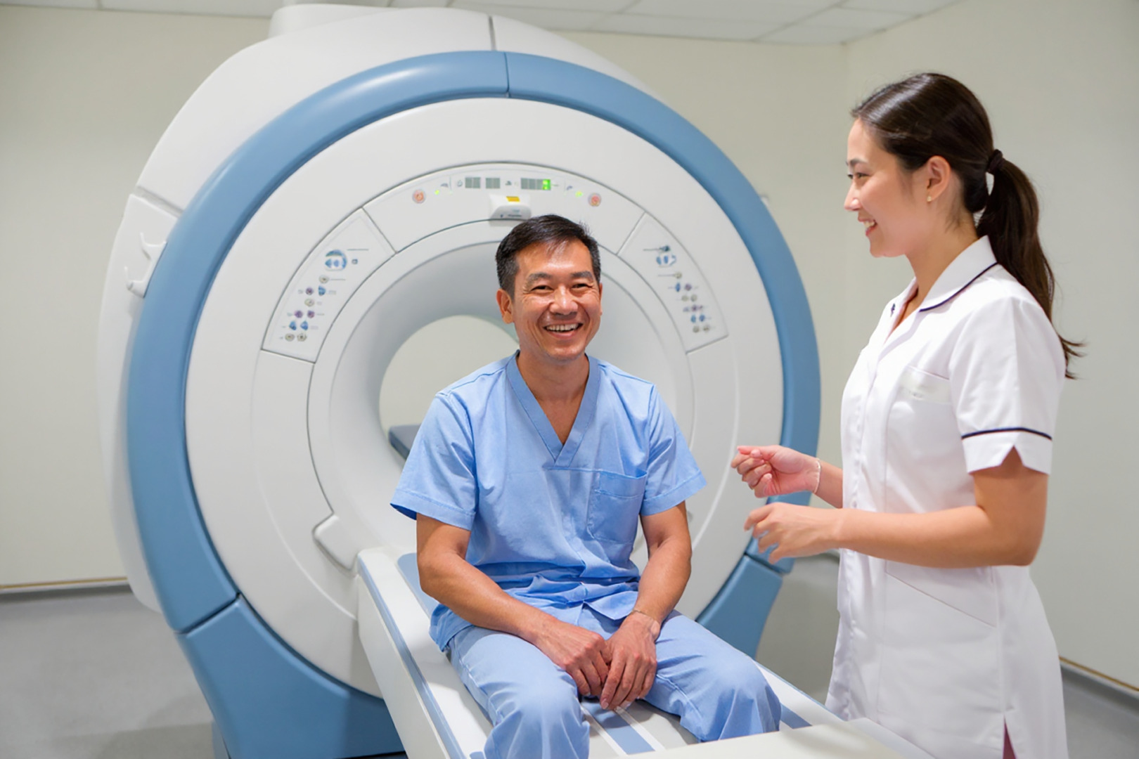
Accurate diagnosis of a chest wall tumour relies on advanced imaging and tissue analysis. Magnetic resonance imaging (MRI) is the gold standard, providing detailed visualisation of the tumour’s extent and infiltration into surrounding soft tissues.
A CT scan is also a key diagnostic tool for evaluating chest wall tumours. It provides valuable information on tumour size, location and bone involvement. This will aid with surgical planning if necessary. Contrast enhancement on CT scans also helps assess tumour margins and vascularity, allowing better differentiation between tumour tissue and surrounding structures. Contrast-enhanced imaging also requires a trained expert to distinguish desmoid tumours from sarcomas or benign fibromas.
A biopsy, either core needle or open, is typically performed to confirm the diagnosis.
Monitoring with Active Surveillance
For asymptomatic or stable chest wall desmoid tumours, active surveillance is needed. This strategy involves regular MRI or CT scans to monitor tumour size and behaviour, with follow-up intervals (typically every 6–12 months) tailored to the tumour’s characteristics and growth rate. A multidisciplinary team of surgeons, radiologists, and pathologists ensures that subtle changes are detected early, enabling timely intervention when needed. Importantly, active surveillance can minimise unnecessary procedures in the future while maintaining a safety net for patients at risk of progression.
Treatment Pathways
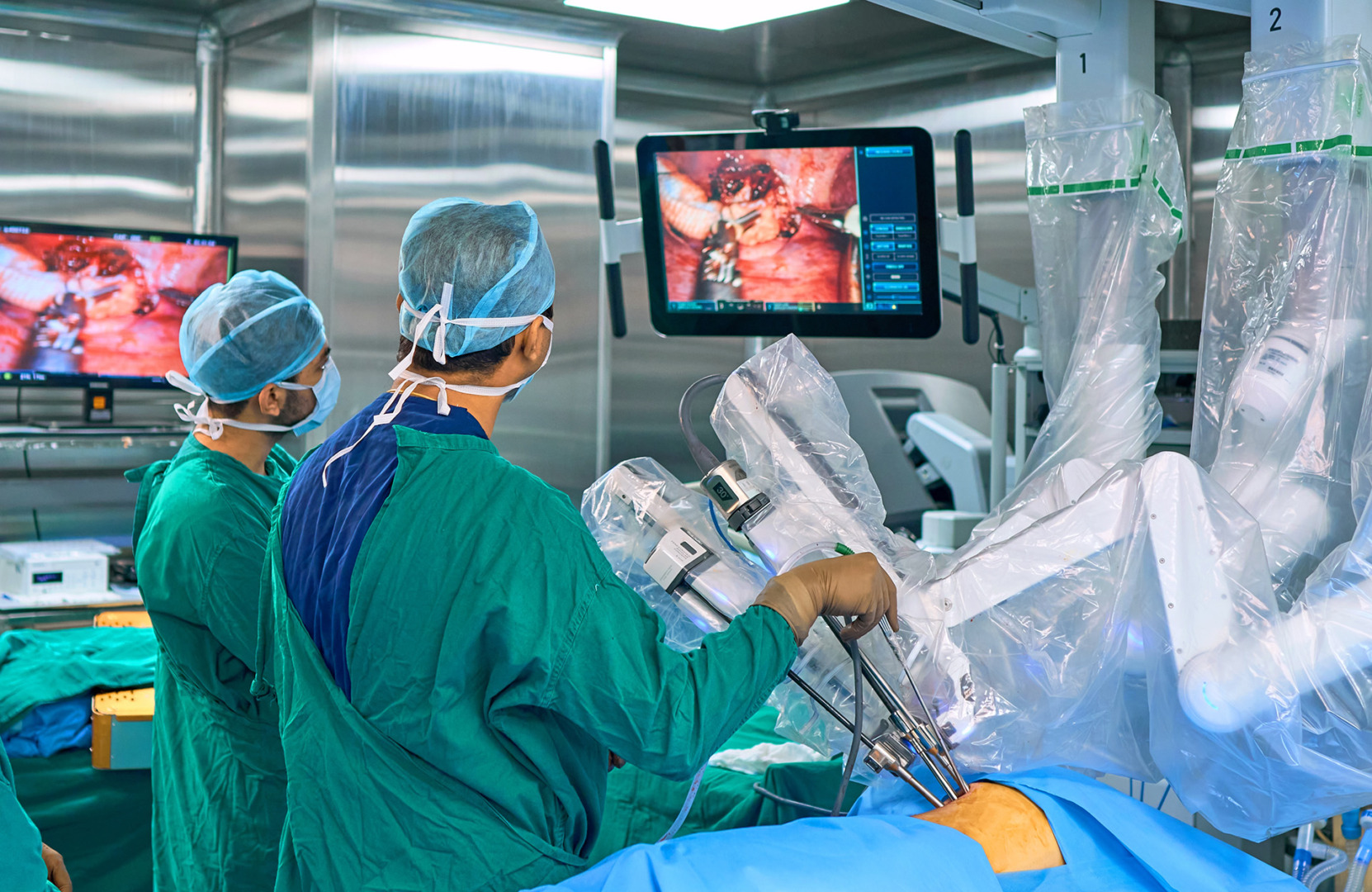
Surgical resection, specifically tumour excision, is a standard treatment for resectable tumours, aiming for complete removal with clear margins to reduce the risk of recurrence. In some cases, a bloc resection involving adjacent structures such as ribs or muscles is necessary. This often requires reconstruction with meshes or grafts to maintain chest wall integrity.
Radiation therapy may be used post-surgery or for inoperable tumours. Targeted therapies, such as tyrosine kinase inhibitors (e.g., imatinib, sorafenib) or gamma-secretase inhibitors (e.g., nirogacestat), show promise for unresectable or recurrent tumours. Hormonal therapies, such as tamoxifen, are selectively used in estrogen receptor-positive cases. Active surveillance is often favoured for stable tumours to avoid unnecessary interventions in future.
Outlook and Recurrence Risks
The prognosis for chest wall desmoid tumours varies, with recurrence rates ranging from 20 to 40% post-surgery. Factors such as tumour size, location and margin status during resection significantly influence outcomes. Larger or incompletely removed tumours carry a higher risk of recurrence, so they require long-term follow-up with periodic imaging to detect regrowth early, enabling timely adjustments to care plans and improving long-term outcomes.
When to Schedule a Consultation
Chest wall desmoid tumours demand early diagnosis and personalised treatments to manage their aggressive behaviour effectively. From active surveillance to advanced surgical techniques and emerging therapies, treatment options continue to expand.
If you’re experiencing symptoms or have a recent diagnosis of a desmoid tumour, consult a specialist. For personalised guidance, schedule a consultation with some of Singapore’s best professionals on desmoid tumours at the Neumark Lung & Chest Surgery Centre.