lung nodule treatments in Singapore

Lung Nodule Treatment in Singapore

A lung nodule (or pulmonary nodule) is a small, round growth typically seen as spots on the chest X-ray or CT scan of the lungs. Most nodules are benign. However, a careful clinical evaluation is necessary to assess the risk of malignancy, which could save your life. Despite the low chances of lung nodules being cancerous, seeing a thoracic specialist to differentiate between benign and malignant lung nodules as early as possible is essential.

Lung nodules are small (under 3cm) lung spots, usually benign, often found incidentally on imaging, and typically caused by prior infection or inflammation. While rarely cancerous, most are asymptomatic, though large ones may cause coughing or shortness of breath. Diagnosis of lung nodules involves imaging (X-ray, CT), and management includes watchful waiting, biopsy, or treatment based on the nodule’s characteristics and the patient’s risk profile. Most lung nodules are non-malignant and can be left alone.

Discovering a lung nodule on a chest X-ray or CT scan is concerning, but understanding these growths and the available treatment options provides reassurance. As lung specialists, Neumark Lung & Chest Surgical Centre guides patients through the whole process from diagnosis to management of lung nodules.

SG lung nodule clinic

What Exactly Are Lung Nodules?

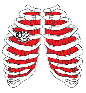
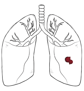
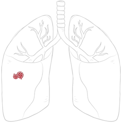
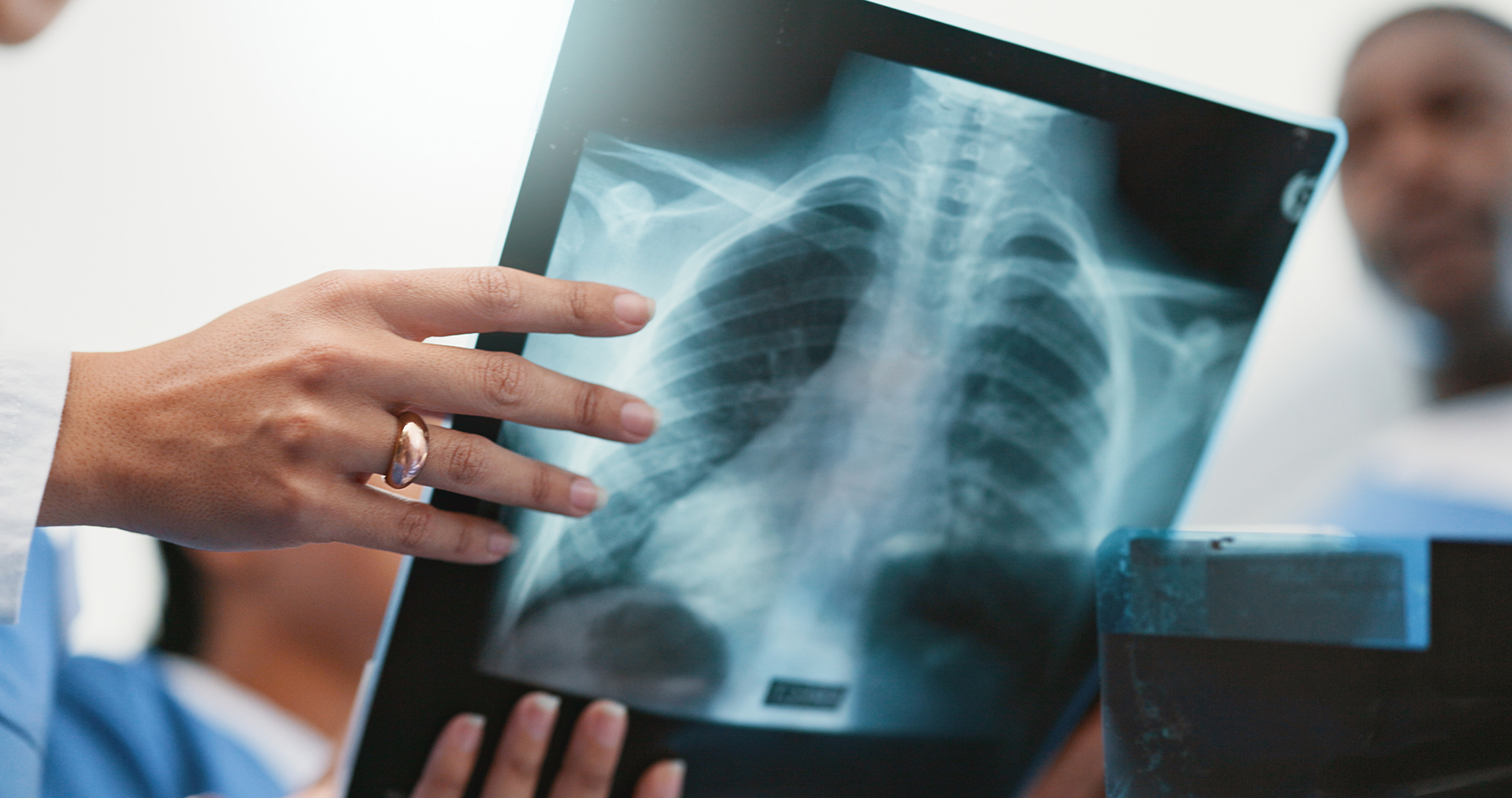
A pulmonary nodule or a lung nodule is a small, typically benign, round or oval tissue growth seen on imaging. Having several small white spots on the lungs (nodules) can indicate prior infection or irritation. Common in people over 50, most are less than 3cm in diameter, and about 95% are non-cancerous. A lung nodule is often detected incidentally during unrelated imaging, facilitating early monitoring. 


Lung Nodules Terminology

Understanding the specific terminology in your radiology report describing your pulmonary nodule can help you grasp your diagnosis.

  • Solid nodules, seen as completely opaque spots on CT scans, are the most common type and are often benign. Their evaluation depends on the nodule’s size and other factors.
  • Ground-glass nodules (GGOs) are hazy lung opacities through which underlying lung structures are visible. They may signal early or slow-growing changes, often needing long-term monitoring.
  • Part-solid nodules (subsolid nodules) have both solid and ground-glass components. The solid part requires closer attention as it may indicate significant changes.


Calcified vs Non-Calcified Nodules

Your report may distinguish whether a nodule is calcified or non-calcified.

Calcified nodules appear bright on CT scans and are often the result of a previous infection, such as tuberculosis or a fungal infection, that has healed. Certain patterns of calcification, like a completely calcified nodule or one with calcification in the centre, are reassuring signs that the nodule is almost certainly benign. Non-calcified nodules do not contain these calcium deposits. Most soft, non-bony lumps are harmless but should be evaluated by a physician.


nodule on lung


Importance of Early Lung Nodule Screening

Lung cancer is Singapore’s third most common cancer. Early detection and removal are critical for survival, so doctors treat every small lung spot as a potential cancer to ensure optimal patient outcome and ease worry about your lung nodule.

This approach, whilst it might seem overly cautious, has proven to be life-saving for many patients. Early screening for potentially cancerous nodules improves patient outcomes through timely treatment.


Causes, Risk Factors and Lung Nodule Symptoms

Most lung nodules are asymptomatic and found incidentally. Possible symptoms of lung nodules include chest pain, shortness of breath, coughing up blood, fatigue, fever, unexplained weight loss, or wheezing. Symptoms alone do not confirm cancer or guarantee a benign nodule; professional evaluation is necessary.

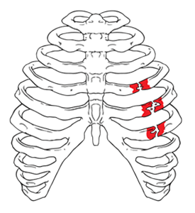
Sometimes, if a small lump is near a breathing tube, it can compress or partially block it. This can cause respiratory difficulties or recurrent infections. If you have breathing problems that don’t go away, tell your physician.

When to seek urgent care: If you experience coughing up blood, sudden, severe breathlessness, or acute, rapidly worsening chest pain, seek medical attention promptly.


nodules on the lungs


Sizes of Lung Nodules

It is normal to ask, “What size of lung nodule is worrisome?” because lung nodule size is a key indicator of malignancy risk.


Size Risk Factor
< 6 mm:  Very low risk (typically < 1%)
6-8 mm Slightly higher risk (around 0.5% to 2%)
8-20 mm Significantly increased risk (5% to 15%, depending on other factors)
> 20 mm (2 cm) Highly concerning; cancer rates can exceed 50% in high-risk individuals
> 3 cm Classified as a lung mass, usually requiring immediate investigation


However, size is not the only factor. Nodule shape, density, border regularity, and temporal changes are crucial. Patient risk factors like age, smoking history and family cancer history also influence how worrisome a nodule of any size may be.


lung nodule symptoms

Advanced Detection of Lung Nodules and Follow-up

The Neumark Lung Screening Programme uses a quick, 10-minute low-dose CT scan (a special, low-radiation X-ray) to detect small nodules. This scan is better than a regular chest X-ray and can find spots as tiny as 2-3mm.

Finding these lung spots is common when people undergo low-dose CT scans or even regular X-rays for other respiratory problems. A multidisciplinary care team reviews all results to assess the nodule’s size, shape, consistency, and location, thereby informing treatment and management options.

Once your scan results are available, you will meet with our pulmonologist. They will review the results with you and answer any questions. In this meeting, they will present the findings, discuss any personal risk factors you may have, and recommend the most appropriate approach for monitoring or treating the nodule.

What Happens Next? A Step-by-Step Explanation

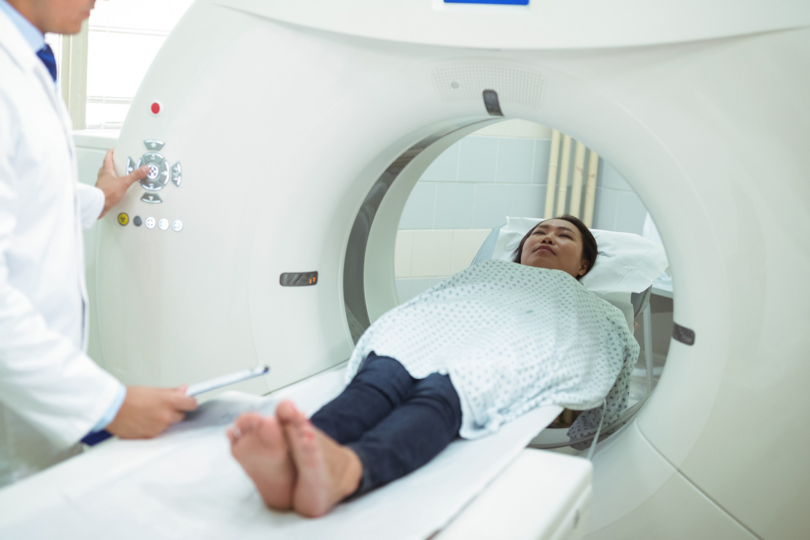
After your CT scan, the following steps help determine the best course of action:

  1. Radiologist report review and risk estimation: We assess your overall risk from the scan based on the spot’s size and appearance (solid, ground-glass, or mixed), as well as patient factors such as age and smoking history.
  2. Additional imaging with PET-CT (when appropriate): For larger or more concerning nodules, PET-CT may be indicated to assess cellular activity. However, lung infections or swelling can also look active, and small or slow-growing cancers might be missed. 
  3. Tests for infection or inflammation: If there’s reason to suspect an infectious or inflammatory cause, additional imaging tests, such as blood tests, sputum cultures, or other investigations, may be ordered to help clarify the diagnosis.
  4. Decision: Based on all gathered information, the most appropriate next step is determined, whether it is surveillance, biopsy, or removal. Low-risk nodules are monitored with follow-up scans. Higher-risk nodules may require a biopsy to obtain a tissue sample. Surgery might be recommended for highly suspicious nodules or when biopsy isn’t feasible.


How Long Does Monitoring Last?

Stable solid lung nodules that have not grown for approximately 2 years have a significantly reduced cancer risk, often eliminating the need for further imaging. Ground-glass and part-solid nodules, however, may require longer monitoring due to their slow rate of change. Follow-up uses low-dose CT to minimise radiation while tracking changes.


lung nodule detection


Do I Need a Biopsy, and How Is It Done?

If the first scan shows a spot in the lung, doctors usually obtain a small tissue sample via a lung biopsy to examine it under a microscope. This is the most definitive method for determining whether the nodule is benign or malignant.


When Is a Biopsy More Likely?

A lung biopsy might be advised for pulmonary nodules if they: 

  • Grow on follow-up imaging
  • Look suspicious on a CT scan (e.g. irregular borders, solid parts)
  • Are large (typically 8mm or more)
  • Have a high-risk history (like heavy smoking or previous cancer)

Very small nodules are usually monitored. A biopsy is typically considered only if the nodule grows or changes, as it can be hard to sample them safely and accurately.


Biopsy Approaches

The optimal tissue sampling method depends on the nodule’s location and characteristics.

  • CT-guided needle biopsy: For peripheral lung nodules, a thin needle is advanced through the chest wall under CT guidance to obtain a precise specimen. This day procedure typically uses local anaesthesia, which your specialist will discuss with you.
  • Bronchoscopy-based biopsy: Used for nodules near airways or central lung areas, a bronchoscope (a flexible tube with a camera) is guided via the mouth or nose, using advanced navigation, to reach hard-to-access nodules.
  • Surgical biopsy (VATS or U-VATS wedge resection): For highly suspicious nodules or after failed biopsies, this minimally invasive procedure provides a combined diagnostic and therapeutic approach to the removal of lung nodules.

Your pulmonologist will discuss the most appropriate approach, including its benefits and potential risks.


lung nodule surgery


Lung Nodule Treatment Options

If you get a diagnosis of a lung nodule, you’ll need to see a thoracic surgeon. The next treatment depends on factors such as the nodule’s size, location, appearance, and the patient’s overall health.


Non-Surgical Management Options

Not all lung nodules need surgery. Non-surgical options may be suitable depending on the diagnosis. Here are some options:

  • Active surveillance: Low-risk, small nodules are often monitored with regular follow-up CT scans to track changes and prevent unnecessary procedures.
  • Antibiotic or antifungal treatment: For suspected or confirmed bacterial/fungal infections, medication will be prescribed, with follow-up imaging used to confirm that the nodule resolves with treatment.
  • Treatment of underlying inflammatory conditions: Nodules from inflammatory conditions (e.g., sarcoidosis, rheumatoid arthritis) require coordinated management with a specialist to address the underlying cause.


Surgical Treatment Options

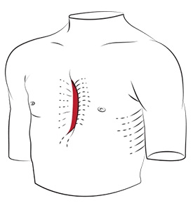
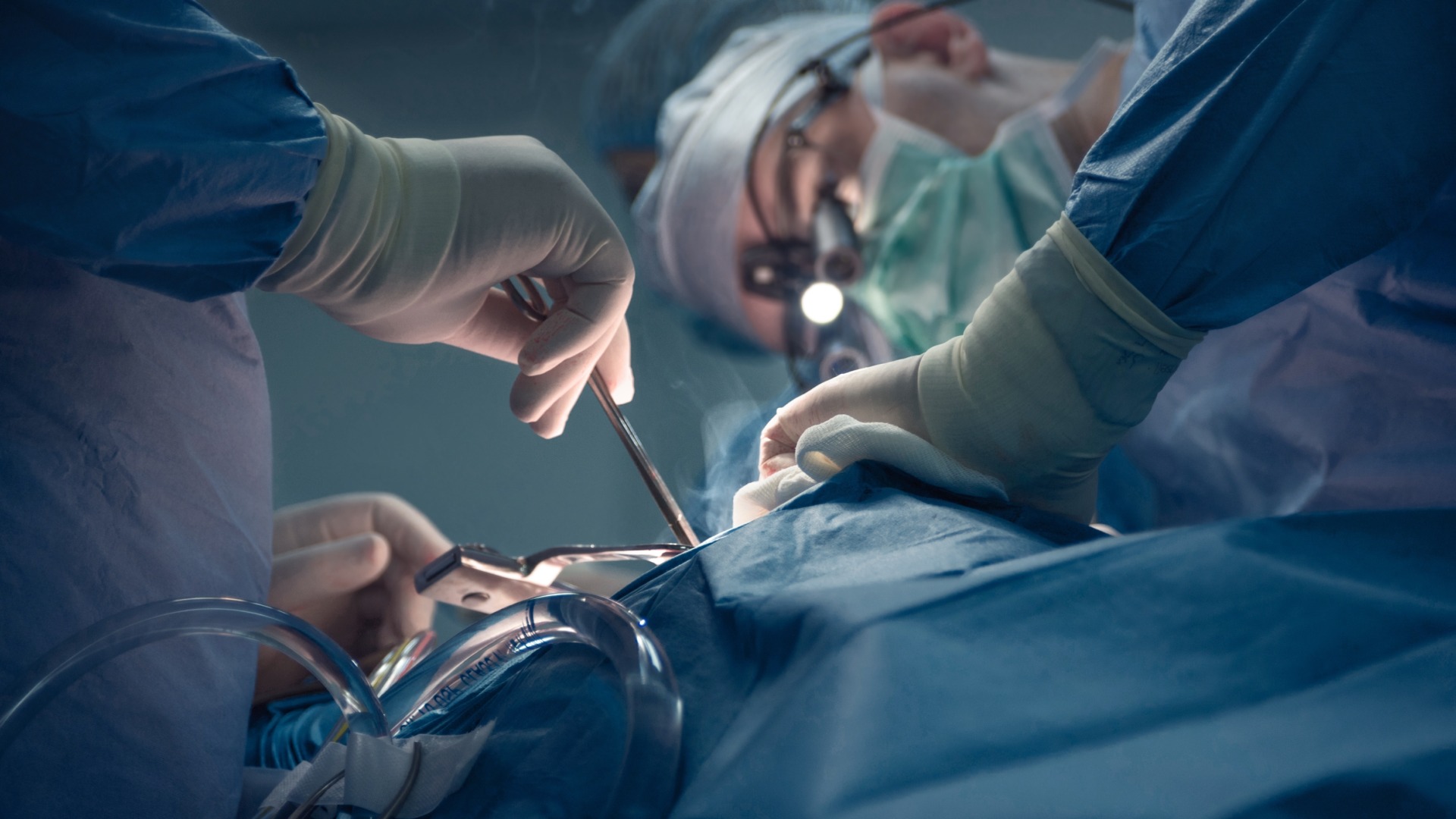
Neumark’s surgical approaches represent the state of the art in the removal of lung nodules. Our minimally invasive options provide excellent outcomes with less discomfort and quicker recovery. Wedge resection is an operation in which a small, wedge-shaped piece of the lung containing the abnormal lump is removed. To do this, we perform either:
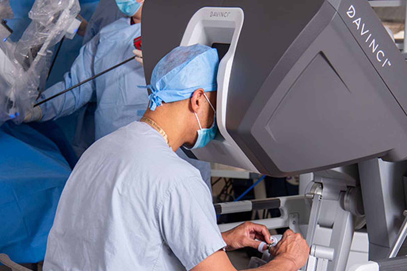
 

Robotic Thoracic Surgery Uniportal Video-Assisted Thoracoscopic Surgery
Our skilled surgeons use the da Vinci Robotic Thoracic Surgery (RATS) system for chest operations. This advanced robot provides the surgeon with better views and greater control, enabling them to remove lung nodules with greater accuracy while preserving as much healthy lung tissue as possible. Uniportal Video-Assisted Thoracic Surgery (U-VATS) is a technique for removing lung nodules through a single small incision. This means less pain, less scarring, and a quick return to normal life, all while successfully removing the problem area. 


lung nodule removal surgery


Treatment for Cancerous Lung Nodules

What Does It Mean If My Nodule Is Cancerous?

Confirmed lung nodules are often early-stage. Next steps include staging scans (e.g., PET and brain imaging) to assess for spread. A multidisciplinary care team of surgeons, oncologists and radiologists will develop a personalised treatment plan. For patients unfit for surgery, highly focused stereotactic body radiation therapy (SBRT) may be an effective alternative. 


Surgical Treatment for Lung Cancer

Treating lung nodules that are cancerous requires a comprehensive, personalised approach. The extent depends on the nodule’s size, location, stage, and the patient’s health. Early-stage lung cancers may only require limited resections like wedge resection or segmentectomy to spare healthy lung tissue. Treatment options include minimally invasive surgery, advanced clinical therapies, and, for more advanced cases, a lobectomy. We also partner with medical oncologists for necessary chemotherapy or radiation therapy.


Recovery and Long-term Outcomes

The rate of recovery depends on the type of surgery. If you had a less invasive surgery, you can return to your regular activities in about 2 to 4 weeks, but it may take several months to fully heal internally. We provide comprehensive care after your operation, including managing your pain, guiding you through breathing exercises, gradually increasing your physical activity, arranging home care, and monitoring for complications. Ongoing surveillance is crucial for early detection of lung cancer recurrence.


lung nodule recovery


Why Choose Specialised Care?

At Neumark Lung & Chest Surgical Centre, our expert care team manages abnormal lung growths and a range of lung and chest conditions using advanced techniques. Our thoracic surgeons are highly trained in chest surgery and manage both benign and malignant conditions to provide optimal, effective care. Dr Harish Mithiran, Director and Senior Consultant Thoracic Surgeon at Gleneagles Hospital, offers patients expert, personalised care with groundbreaking minimally invasive treatments to improve outcomes. Please call us and schedule your appointment today.


lung nodule specialist

FAQs about Lung Nodules

CT scans detail nodule size, shape, density, and location to estimate cancer risk, but cannot definitively diagnose it. PET-CT may provide additional metabolic information, though it also has limitations.

Ground-glass opacity (GGO) on a CT scan appears hazy, allowing underlying lung markings to be seen, unlike solid nodules. GGOs often need careful long-term monitoring, as some may be slow-growing abnormalities.

Monitoring for stable solid nodules often stops after about 2 years. Ground-glass and part-solid nodules require longer follow-up, possibly several years, due to slow nodular changes. Your care team will tailor the schedule based on your risk factors and successive scan results.

A PET-CT scan is best for evaluating larger, uncertain nodules for cancer by detecting increased metabolic activity. However, it has limitations: inflammation can lead to false positives, and very small or slow-growing cancers can yield false negatives. A doctor will recommend a PET-CT scan when its utility is high for the specific case.

Lung nodule cancer risk correlates with size: under 6mm is very low (<1%); 6-8mm is 0.5-2%; 8-20mm is 5-15%; and over 20mm can exceed 50% in high-risk patients. However, a nodule’s appearance, growth, and personal risk factors are also crucial in determining the level of concern.

Pulmonary nodules are very common, found in up to 50% of adults undergoing chest CT scans, especially those over 50. The vast majority (around 95%) are benign. This high detection rate is due to the sensitivity of modern CT scanners, which can detect nodules as small as a few millimetres.

Lung nodules can shrink or disappear, often if caused by resolving infection or inflammation. Follow-up imaging is essential to observe this behaviour. A shrinking or resolving nodule is reassuring; however, further follow-up may still be warranted.

DISCLAIMER: The information provided on this website is for general informational purposes only and is not intended as a substitute for professional medical advice, diagnosis, or treatment. The use of this website does not create a doctor-patient relationship and no medical advice should be inferred or assumed. It is the user’s sole responsibility to seek the advice of their healthcare professionals for any medical concerns they may have and the user should not disregard, or delay, prompt medical advice for any such condition.

Lung Nodule Treatment in Singapore

Neumark Lung and Chest Surgery Centre benefits from the expertise of a multidisciplinary team led by Dr Harish Mithiran, senior consulting thoracic surgeon at Gleneagles Hospital and Mt Alvernia Hospital.

Neumark is a lung and chest specialist centre with access to leading treatment modalities to achieve the best possible outcomes for lung disease and preventative patient screening.

Our foremost priority is to treat your condition as effectively as possible. Schedule a private consultation today; complete the form below, call, +65 6908 2145; WhatsApp, +65 9726 2485; or email, info@neumarksurgery.com.