

When you get news of a possible lung nodule, anxiety may naturally follow. These small lesions near the lung’s outer surface present unique challenges and opportunities for diagnosis and treatment. Fortunately, advanced treatment options for lung nodules are available with expert care. A subpleural node does not have to be something you go through alone, so make sure you have the right expert team by your side.
What Is A Subpleural Nodule In The Lung?
A subpleural nodule is a small, round or oval lesion within 2cm of the pleural surface. These nodules are 3 to 30 mm in diameter, although size doesn’t determine clinical significance. On CT scans, they appear as well-defined or irregularly bordered masses against surrounding lung tissue. Subpleural nodules can be a solitary pulmonary nodule, which is a single well-circumscribed lesion without associated lymphadenopathy. Common locations for subpleural pulmonary nodes are the upper and lower lobe periphery, where they are most accessible for imaging and surgery.
Density characteristics vary significantly depending on the underlying cause and whether the condition is benign or malignant. Benign nodules have smooth, well-defined borders and consistent density. Malignant lesions typically exhibit spiculated margins, heterogeneous density patterns, or surrounding ground-glass changes, indicating active disease processes. These features are best visualised on CT images, and nodules identified on CT scans are evaluated using specific protocols to guide further management.
Causes and Risk Factors
Many things can cause subpleural nodules. Most are harmless and arise from past infections, such as tuberculosis, fungal infections or pneumonia, which can leave behind small scars or areas of inflammation (granulomas). Some individuals may also develop small, harmless growths, such as hamartomas or fibrotic nodules, as their lungs heal from prior inflammation or exposure to irritants. Inflammatory conditions such as sarcoidosis or rheumatoid arthritis can also produce nodules similar to those seen in sarcoidosis.
On the other hand, subpleural nodules can also be cancerous. They may be early-stage primary lung cancers, especially adenocarcinomas that develop in the outer parts of the lung. In some cases, the nodules may be signs of cancer that have spread to the lungs from other areas like the breast, colon or kidneys.
Several risk factors increase the likelihood of a subpleural nodule being malignant. People who smoke or have smoked in the past are at a much higher risk, even many years after quitting. Age is another important factor, as the risk of cancer increases after 50. Exposure to harmful substances, such as asbestos, radon and industrial chemicals, can also increase the risk of developing these conditions. Those with chronic lung conditions like COPD, pulmonary fibrosis or a history of tuberculosis may also develop subpleural nodules as part of these diseases.
In short, while most subpleural nodules are harmless, certain factors like smoking, age, past lung infections or a personal or family history of cancer make it important to take them seriously and evaluate them thoroughly.
Symptoms and When to Worry
Most subpleural nodules are asymptomatic and are discovered incidentally. This asymptomatic nature is why they are often found incidentally during routine imaging and why screening is important in high-risk populations.
When symptoms occur, the nodules usually reflect the underlying cause rather than the nodules themselves. Localised chest pain sometimes suggests pleural involvement or rapid growth affecting surrounding structures.
Unexplained weight loss, fatigue or decreased exercise tolerance may accompany malignant subpleural nodules, especially primary lung cancer or metastatic disease. These constitutional symptoms develop gradually and are often overlooked initially.
Red flags for urgent evaluation include rapid nodule growth on serial imaging, new symptom development in previously asymptomatic patients, or the appearance of multiple new nodules. Changes in nodule characteristics, such as increasing density, irregular borders, or surrounding inflammatory changes, require prompt specialist assessment.

Imaging and Diagnosis of Subpleural Nodules
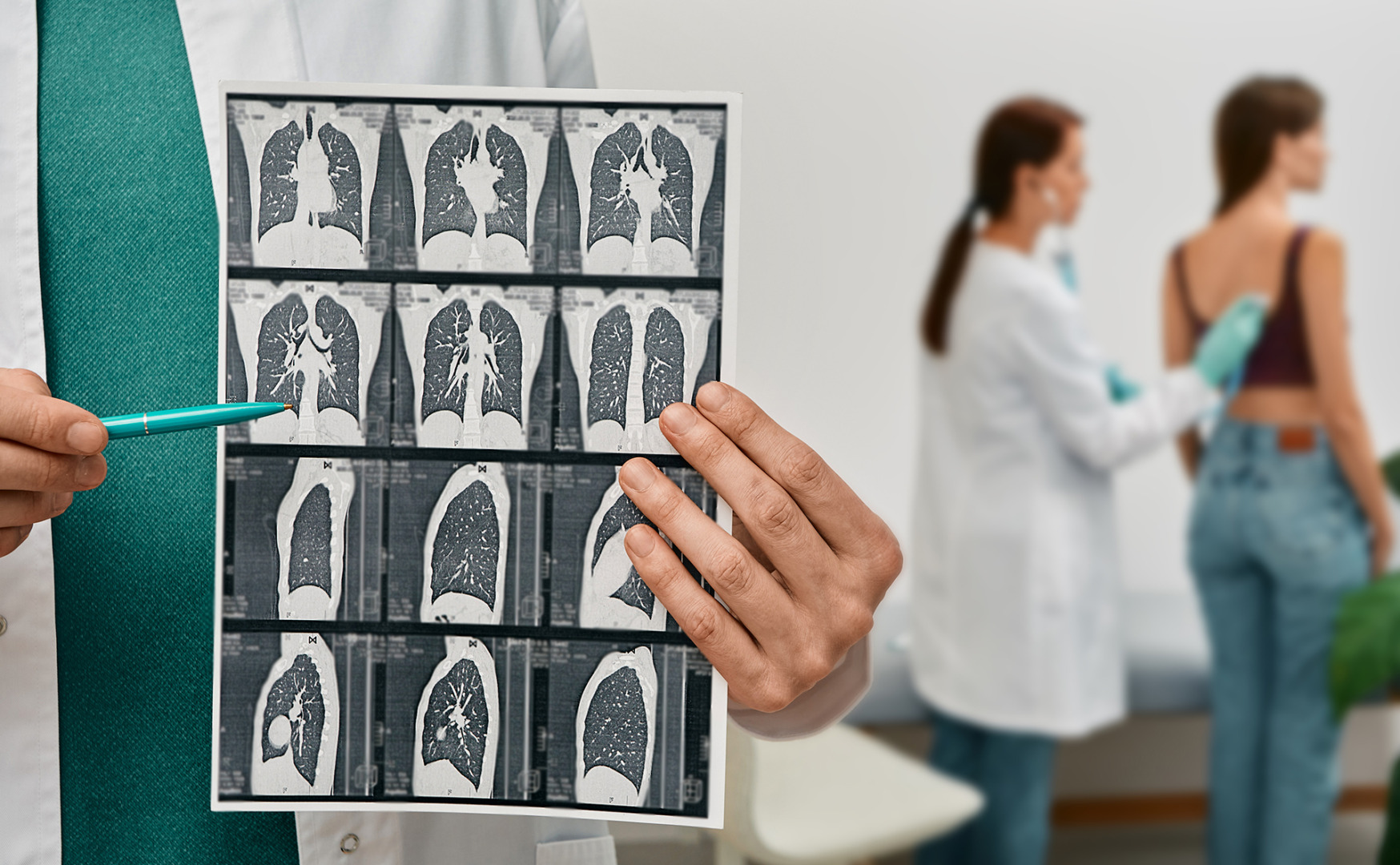
Modern imaging plays a key role in evaluating subpleural nodules. A low-dose CT scan is the first and most important step. It provides detailed images of the lungs while minimising radiation exposure. Special high-resolution CT scans can reveal the size, shape, borders, and density of the nodule, as well as its relationship to surrounding lung tissue. Comparing current and previous scans helps to spot any growth or changes over time.
Low-dose CT (LDCT) lung cancer screening has changed how subpleural nodules are detected, often identifying them in high-risk people before symptoms appear. Unlike chest X-rays, which can miss small nodules, CT scans are much more sensitive, especially in early-stage disease.
These screening programmes target people aged 50-80 who have smoked heavily for many years. Modern CT scans are so detailed that they often detect many small nodules, most of which are not cancerous. Both lungs should be carefully assessed to detect any signs of disease.
Certain CT scan features can suggest whether a nodule is likely benign or cancerous. For example, calcification within a nodule often indicates an old infection, and spiculated or irregular edges raise concern for cancer. Nodules with ground-glass changes or uneven density may indicate inflammation or malignancy.
PET-CT scans add another layer by showing how active the nodule is. Cancer tends to utilise more glucose, which appears as a bright spot on the scan. However, inflammation can also increase uptake, so PET scans are most helpful when the nodule is large or unclear on CT alone.
Contrast-enhanced CT scans can help distinguish between infection, inflammation and cancer based on how the nodule takes up contrast. Radiologists combine all this imaging information with your medical history to decide the next steps.
If a nodule is larger than 8mm, has irregular borders, is growing or is suspicious on imaging, a biopsy may be recommended.
Subpleural Nodule vs Other Lung Nodules
Subpleural nodules sit at the outer edge of the lung near the chest wall. This makes them easier to see on scans and often simpler to access for surgery or biopsy. Unlike nodules near the airways, subpleural nodules rarely cause symptoms like cough or shortness of breath unless they grow large or affect the pleura.
Risk assessment for subpleural nodules follows the same guidelines as for other lung nodules, but the location can influence decision-making. Lung cancers like adenocarcinoma often appear in subpleural areas, so doctors pay close attention to size, shape and growth when evaluating them.
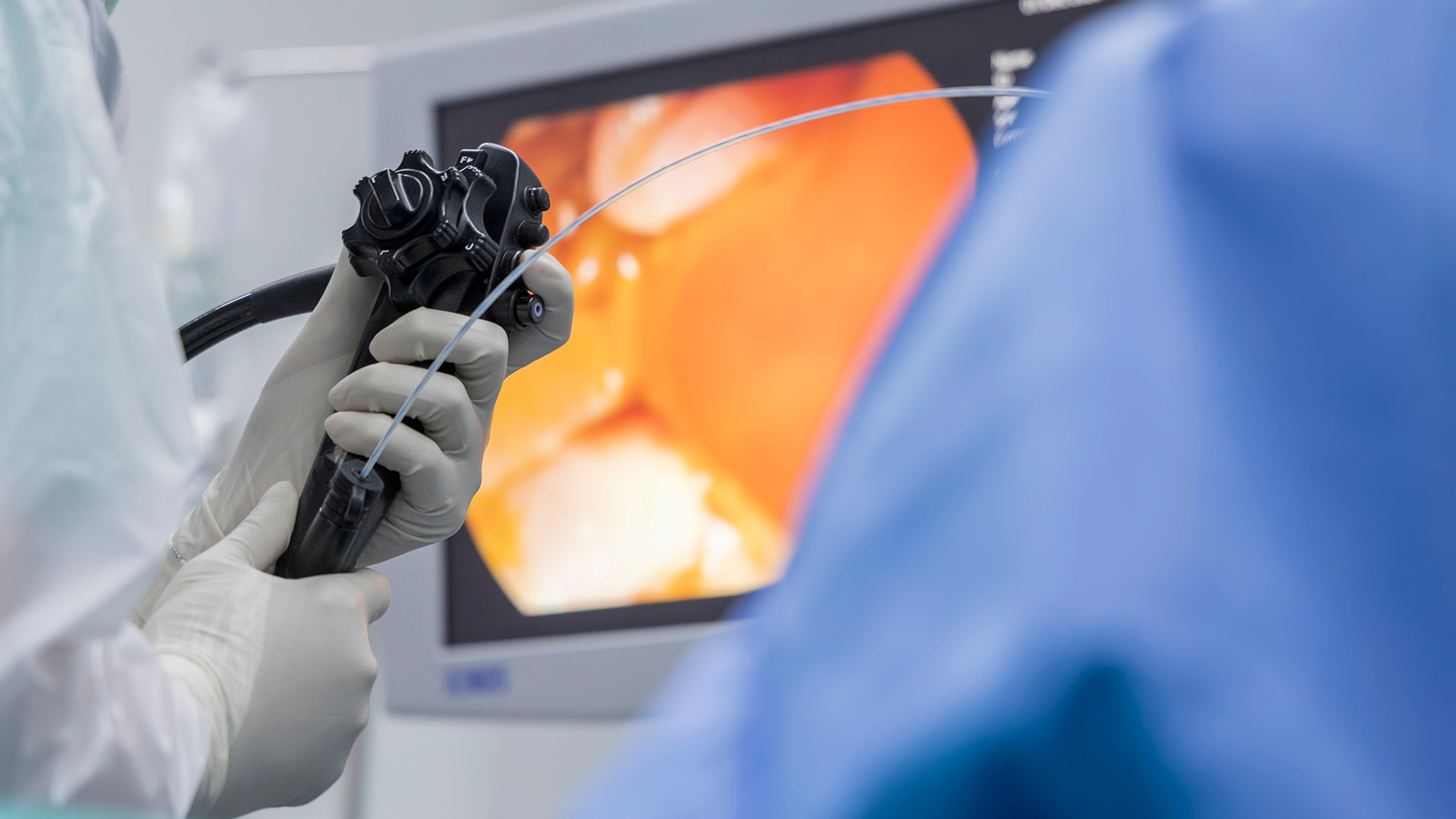
As subpleural nodules are located near the chest wall, they are well-suited for CT-guided needle biopsies, which eliminates the need for more invasive bronchoscopic procedures. However, this approach carries a slightly higher risk of pneumothorax due to its proximity to the pleural space.
Managing these nodules effectively often involves a team of specialists working together to assess risk and decide the best course of action.

Management and Treatment Options
Management of a subpleural nodule depends on its cancer risk, the patient’s health and their personal preferences. For many small nodules with low-risk features, the best first step is watchful waiting. This involves follow-up CT scans every 3-6 months with longer intervals if the nodule remains stable. If it shows no growth over 2 years, monitoring may be reduced or stopped.
Surgery is considered when a nodule grows, changes shape, or becomes large enough to raise concern. The decision also depends on the patient’s age, health and whether they prefer a clear diagnosis over continued monitoring.
Video-assisted thoracoscopic surgery (VATS) is the gold standard for removing subpleural nodules. It uses small incisions and special tools to remove the nodule with minimal trauma. Most of these nodules can be treated with a wedge resection, which involves removing the growth with a small margin of healthy lung tissue. In more complex cases, robotic-assisted surgery may be used. This offers more precision, especially when multiple nodules are present or delicate dissection is needed.
Removing a cancerous nodule early gives patients the best chance of a cure. For example, surgery for stage I lung cancer can provide 5-year survival rates over 80%. Even if the nodule turns out to be benign, the reassurance of a confirmed diagnosis often outweighs the small risks of minimally invasive surgery.
Prognosis and Follow-Up
Outcomes for people with subpleural nodules depend on the cause, but most have excellent outcomes. Importantly, benign nodules typically require no treatment beyond regular scans and rarely impact daily life.
If the nodule is cancerous and caught early, surgery often gives excellent results. Small cancers near the lung edge, when removed before they spread to the lymph node, can have 5-year survival rates of over 90%.
Follow-up plans depend on how the nodule initially appeared. Low-risk nodules that remain stable over time may only need annual scans. After surgery, regular imaging helps check for recurrence, though scan intervals typically lengthen over time.
Quitting smoking is key to long-term health. Staying active, avoiding lung irritants, getting vaccinated, and managing any chronic lung disease can all help protect lung function and reduce the risk of future lung disease.

When To See A Specialist
Nodules that develop symptoms, change shape, or show increased metabolic activity on a PET scan should be evaluated by an experienced thoracic specialist as soon as possible.
Patients with multiple subpleural nodules face complex management decisions and benefit from specialist input. The differential diagnosis for multiple nodules includes metastatic disease, inflammatory conditions and synchronous primary lung cancers, each requiring different management approaches.
At Neumark Lung & Chest Surgery Centre, patients benefit from comprehensive evaluation protocols that incorporate the latest imaging techniques, minimally invasive diagnostic procedures, and advanced surgical options. The combination of specialist expertise and state-of-the-art technology ensures that patients receive care tailored to their individual needs.
Contact Neumark Lung & Chest Surgery Centre today to book a consultation with our thoracic surgery specialists.