Subpleural nodules are often incidental findings on CT scans, causing understandable anxiety in patients. Their peripheral location can make them difficult to access through traditional bronchoscopic methods, necessitating alternative approaches for diagnosis and treatment.
Significant advancements in the management of lung nodules, particularly subpleural nodules, have made this condition treatable. These small growths, located just beneath the pleura (the membrane covering the lungs), present unique challenges in diagnosis and treatment.
In recent years, two minimally invasive surgical techniques have advanced our approach to subpleural nodules: Robotic-Assisted Thoracic Surgery (RATS) and Video-Assisted Thoracic Surgery (VATS). Both methods offer significant advantages over traditional open thoracotomy, particularly for subpleural lesions.
Small growths raising concern in patients
Subpleural nodules, small growths located beneath the lung’s pleural membrane, often raise concerns among patients despite frequently being incidental findings. These nodules can range from benign to malignant, with causes including infections, inflammation, or tumours.
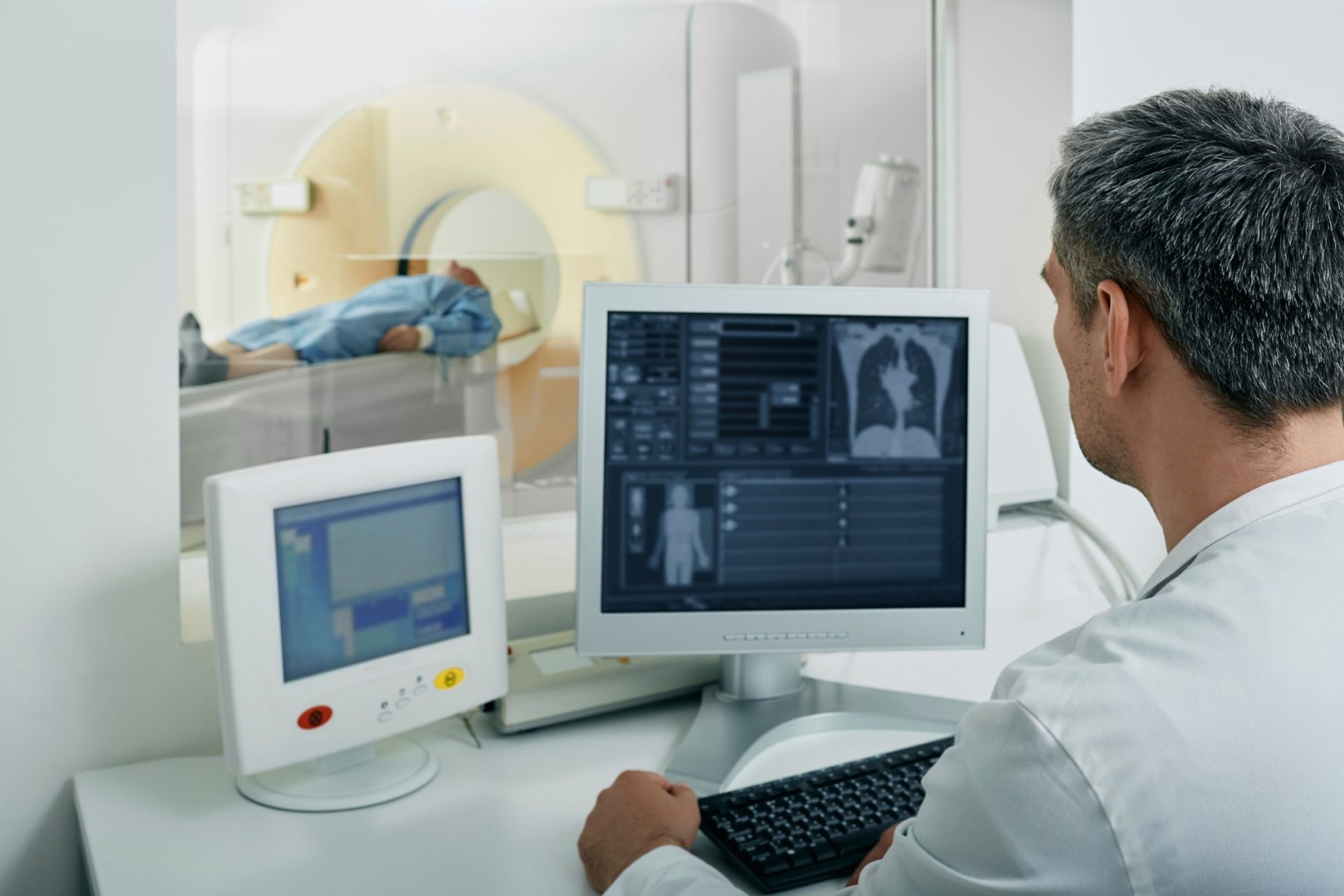
While typically asymptomatic, larger nodules may occasionally cause chest pain or respiratory symptoms. Diagnosis usually involves CT scans, with follow-up imaging or biopsies as needed. The peripheral location of these nodules can complicate diagnosis and treatment, making advanced techniques like RATS and VATS particularly valuable.

Watchful waiting may be appropriate for small, low-risk nodules, while larger or suspicious ones might require surgical intervention or radiation therapy. Patients should understand that many subpleural nodules are benign, but size, growth rate, and appearance influence malignancy risk.
Post-treatment, patients can expect varying recovery times and should be prepared for long-term follow-up. While a diagnosis can cause anxiety, support resources are available. Preventive measures, such as smoking cessation, can also reduce nodule risk.
Ongoing research continues to improve detection and treatment options. By understanding these aspects of subpleural nodules, patients can better navigate their diagnosis and treatment journey, working closely with their healthcare team to determine the most appropriate course of action.
Precision of Robotic Surgery
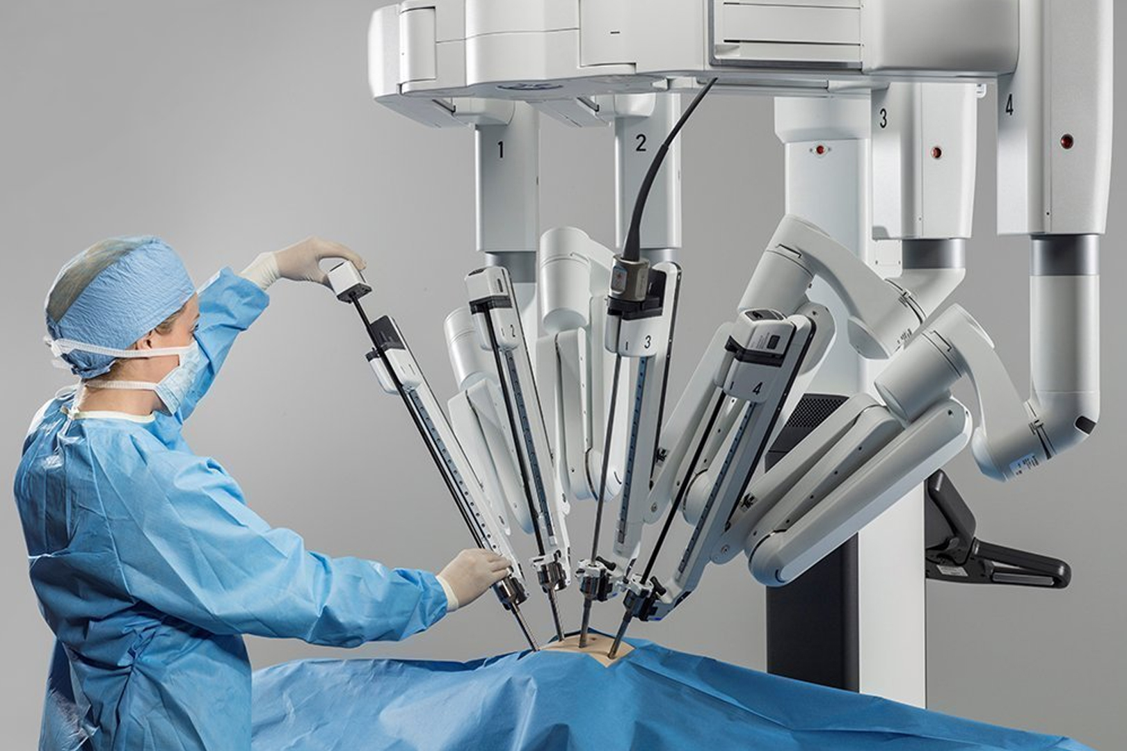
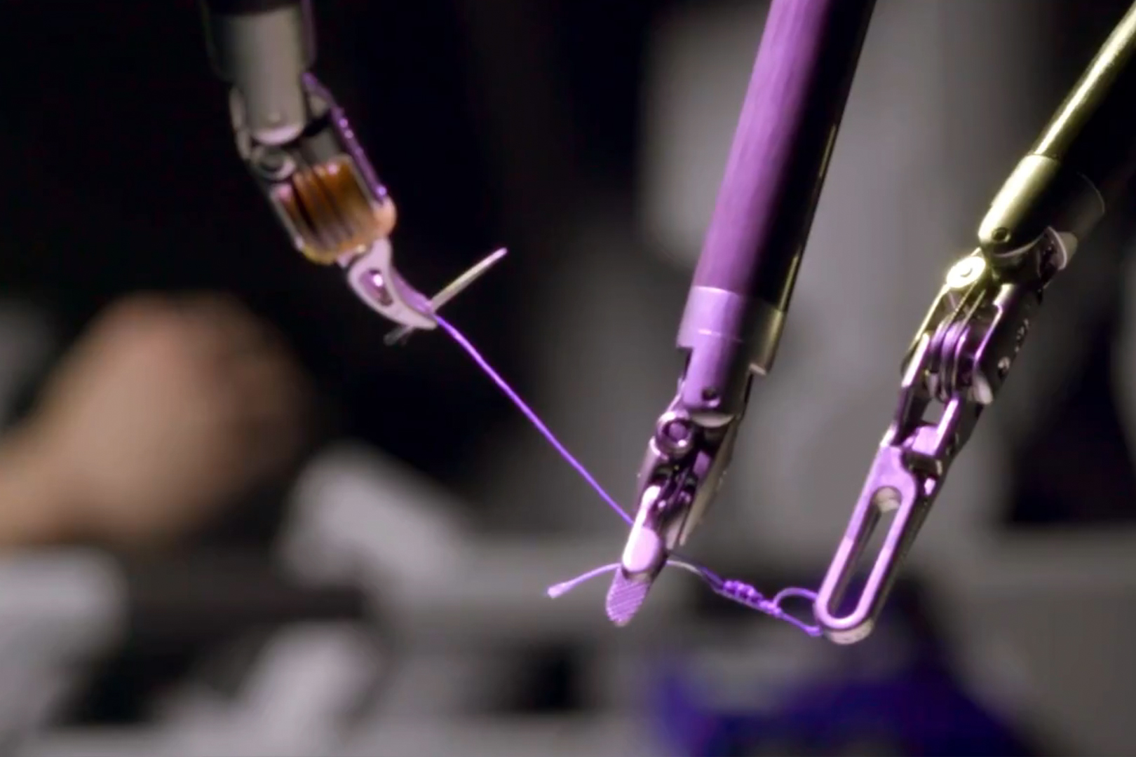
The Robotic-Assisted Thoracic Surgery with the da Vinci system provides unparalleled 3D visualisation and instrument dexterity, allowing for precise dissection and resection of these peripheral subpleural lesions.
The enhanced magnification offered by RATS is particularly beneficial when dealing with subpleural nodules. It allows for clear identification of the nodule and its relationship to surrounding structures, minimising the risk of inadvertent injury to the pleura or adjacent lung tissue.
Moreover, the robot’s articulated instruments can navigate the chest cavity with remarkable agility, reaching areas that might be challenging to access with traditional thoracoscopic instruments. This is especially valuable when dealing with nodules in the posterior or apical regions of the lung.
Minimally Invasive Meets Efficacy with Video-Assisted Surgeries
Video-Assisted Thoracic Surgery, particularly in its uniportal form, offers another excellent option for managing subpleural nodules. The single-incision approach of uniportal VATS results in less postoperative pain and faster recovery compared to multiport techniques.
For subpleural nodules, VATS allows for direct visualisation and palpation of the lung surface, which can be crucial in localising small or ground-glass opacities that might be difficult to identify visually. The technique also permits intraoperative frozen section analysis, enabling immediate decision-making regarding the extent of resection needed.
The minimally invasive nature of VATS translates to shorter hospital stays and quicker return to normal activities for patients. This is particularly beneficial for elderly patients or those with comorbidities who might not tolerate more invasive procedures well.
Choosing Between RATS and VATS
The decision between RATS and VATS for subpleural nodules depends on several factors. Nodule size, exact location, and the patient’s overall health status all make this decision.

“In my practice, I find RATS particularly useful for deeper subpleural nodules or those requiring more complex dissection”, says Dr Harish Mithiran, director of Neumark Lung & Chest Surgery Centre. “VATS, especially uniportal VATS, is often recommended for more superficial lesions or in cases where tactile feedback is crucial. But each patient’s case is unique.”
It’s worth noting that both techniques allow for seamless conversion to open surgery if necessary, ensuring patient safety is never compromised.
“Given the sometimes elusive nature of subpleural nodules, preoperative or intraoperative localisation techniques can be invaluable”, explains Dr Mithiran. Methods such as CT-guided hook wire placement, lipiodol marking, or indocyanine green (ICG) fluorescence have significantly improved a thoracic surgeon’s ability to accurately locate and resect these lesions.
These localisation techniques, combined with the precision of RATS or VATS, have dramatically increased success rates in diagnosing and treating subpleural nodules while minimising tissue loss.
As we continue to refine these techniques and integrate new technologies, the future looks bright for patients facing the challenge of subpleural nodules. If you have been recently diagnosed with a subpleural nodule, consider scheduling a consultation with our lung specialist at Neumark Lung & Chest Surgery Centre.

