

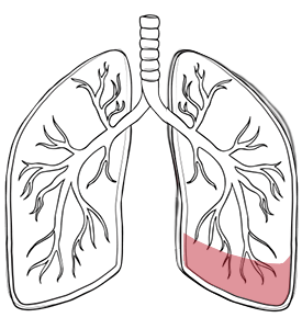
Lung consolidation is an area of the lung that has become filled with something other than air, making it appear denser on imaging. The tiny air sacs, called alveoli, may become filled with pus, blood, inflammatory cells, or other material. On a chest X-ray or a CT scan, this area often appears whiter or denser than the surrounding normal lung.
Lung consolidation is not a diagnosis by itself. Sometimes called pulmonary consolidation, it is a radiological finding, meaning it is something seen on a scan. The important question is why it has happened. In many cases, the condition is caused by pneumonia. In others, it may be linked to fluid in the lungs, bleeding, aspiration, blockage of an air passage, lung cancer, or lung collapse.
The term ‘collapse consolidation’ of the lung may be used when part of the lung has both collapsed and become filled or blocked. This typically happens when an air passage is obstructed by mucus, an infected airway, a foreign body, or, less commonly, a tumour.
Lung consolidation symptoms depend on the cause, size and location of the affected area. Some patients have obvious signs. Others only discover the finding after a chest X-ray or CT scan is done for another reason.
When symptoms do occur, they often include a cough, fever, chest pain, breathlessness, tiredness, and coughing up phlegm. The phlegm may be yellow, green, rust-coloured, or blood-stained, depending on the cause.
Some signs need urgent attention. These include severe breathlessness, sudden or worsening chest pain, coughing up blood, blood in the phlegm, confusion, blue lips, low oxygen levels, or a fever that does not improve. These signs may indicate a serious infectious process or another emergency in the thorax.
Lung consolidation causes include infections, fluid build-ups, bleeding, aspiration, blocked air passages, and cancer.
Pneumonia is the most common cause, and community-acquired pneumonia is the most common type. Bacterial pneumonia is typical and occurs when bacteria, viruses, or other germs infect the lungs and fill the air spaces with inflammatory fluid or pus.
Tuberculosis can also produce consolidation, especially when the symptoms last for weeks or months. The infection may be bilateral, involving both lungs, or limited to the right lung or to a single segment, and a patchy consolidation pattern is common.
Pulmonary oedema is another cause. Excess water builds up in the lungs, typically because the heart is not pumping effectively, raising blood pressure and forcing it out of small blood vessels into the lung’s air sacs.
Pulmonary haemorrhage means that blood replaces the air spaces normally filled by the lung parenchyma.
Aspiration occurs when food, saliva, stomach acid, or vomit enters the lungs instead of moving along the normal route to the stomach. These substances then spread through the airways.
Airway blockage can also lead to consolidation. If mucus or a tumour blocks a bronchus, the affected lung beyond the blockage may collapse or become infected. This is one reason chronic lung consolidation, or consolidation that does not clear, needs further review.
A neoplasm such as lung cancer can sometimes appear as consolidation or cause an infected change behind a blocked airway. Rarely, a secondary deposit from breast cancer, the liver, or another tumour produces a similar appearance.
Other causes include inflammatory lung disease, autoimmune disease, inhalation injury, trauma, and secondary changes after surgery or other secondary causes. The cause is not always obvious from one scan. Doctors usually combine imaging, physical signs, examination findings, and sometimes lab tests or a lung biopsy to determine the cause.
Lung consolidation occurs within the lung tissue, while a pleural effusion is fluid outside the lung, in the space between the lung and the chest wall.
The difference between lung consolidation and pleural effusion is important because the appropriate care differs. In consolidation, the lung parenchyma itself is affected. The air spaces are filled with pus, blood, or inflammatory cells. These inflammatory cells, along with other cells, make the lung appear dense. With an effusion, fluid collects around the lung on the affected side and can press on it from the outside, so the affected side may look opaque.
Both can cause breathlessness, a cough, and chest discomfort. Both can also appear as white areas on a chest radiograph. The effusion may need drainage if the fluid is large, infected, or suspicious for cancer. Lung consolidation usually requires care directed at the underlying cause, such as antibiotics if pneumonia is present, or further testing if cancer or an airway obstruction is suspected.
A person can have both at the same time. For example, pneumonia can cause consolidation and fluid around the lungs. When this happens, lung specialists need to decide whether the main problem is infected lung tissue, fluid on the affected side, or both.
You should see a lung specialist when lung consolidation is severe, persistent, recurrent, unexplained, or linked with worrying signs.
Many cases of pneumonia improve with appropriate care. Follow-up imaging may be recommended to make sure the area clears, especially in older adults, smokers, or patients with risk factors for lung cancer. If the consolidation does not improve, further testing is needed.
Specialist review is important if:
In Singapore, patients often first discover the condition through chest imaging performed for a fever, cough, screening, or a follow-up for another condition.
Lung consolidation is diagnosed with imaging, but the cause is found through clinical assessments and targeted tests.
The pattern, distribution, and overall distribution of lung consolidation, including whether changes are bilateral, guide the differential diagnosis.
Treatment for lung consolidation depends on the cause and may involve medications, supportive care, procedures, or surgery in selected cases.
If pneumonia is the cause, care may include antibiotics, rest, hydration, fever control, and oxygen if needed. Some viral infections need supportive care rather than antibiotics. Severe pneumonia may require hospital care, and pneumonia in older adults can be harder to clear.
If excess lung water is related to heart failure, care may include diuretics and heart medicines, which reduce swelling and fluid accumulation.
If aspiration is the cause, care may involve treating any infected area, improving swallowing safety, managing reflux, and reducing the risk of future aspiration. If bleeding is confirmed, doctors will need to identify and control the source.
When an airway blockage is suspected, care depends on the cause. Mucus plugging may need airway clearance or a bronchoscopy.
A tumour may need a biopsy and cancer treatment.
If consolidation is associated with lung collapse, pneumothorax, the obstructed airway, or trapped lung segment, it must be assessed carefully.
Surgery is not the usual treatment for a simple lung consolidation. It may be considered when there is an unresponsive lung abscess, a suspicious mass, recurrent episodes of infection due to a damaged lung segment, or a complication that cannot be managed with medications or bronchoscopy alone. A thoracic surgeon helps determine whether surgery is needed and whether a minimally invasive approach is appropriate.
Neumark Lung & Chest Surgery Centre helps by identifying the cause of lung consolidation and guiding the appropriate treatment, ranging from medical management to specialist thoracic interventions.
Neumark specialises in minimally invasive thoracic surgery with a multidisciplinary approach led by Dr Harish Mithiran, senior consultant thoracic surgeon at Gleneagles and Mount Alvernia hospitals. For each patient with this condition, this means careful review of scans, signs, prior films, and risk factors before recommending further care.
If you have been told you have lung consolidation or chronic lung consolidation, contact Neumark for a consultation.
DISCLAIMER: The information provided on this website is for general informational purposes only and is not intended as a substitute for professional medical advice, diagnosis, or treatment. The use of this website does not create a doctor-patient relationship and no medical advice should be inferred or assumed. It is the user’s sole responsibility to seek the advice of their healthcare professionals for any medical concerns they may have and the user should not disregard, or delay, prompt medical advice for any such condition.

Neumark Lung & Chest Surgery Centre benefits from the expertise of a multidisciplinary team led by Dr Harish Mithiran, senior consulting thoracic surgeon at Gleneagles Hospital and Mt Alvernia Hospital.
Neumark is a lung and chest specialist centre with access to leading treatment modalities to achieve the best possible outcomes for lung disease and preventative patient screening.
Our foremost priority is to treat your condition as effectively as possible. Schedule a private consultation today; complete the form below, call, +65 6908 2145; WhatsApp, +65 9726 2485; or email, info@neumarksurgery.com.
Gleneagles Medical Centre
6 Napier Road
#02-09 Gleneagles Medical Centre
Singapore 258499
Mount Alvernia Hospital
820 Thomson Road
#06-07 Medical Centre A
Singapore 574623