

Understanding Mediastinal Masses
A mediastinal mass is an abnormal growth in the mediastinum, the middle compartment of the chest between the lungs. This crucial space contains vital structures like the heart, oesophagus, thymus gland, trachea and major blood vessels. Thus, a mass in the mediastinum can be significant even if it is benign.
Mediastinal tumours can be benign or malignant. The anterior mediastinum is the most common site in adults and typically affects middle-aged and older people. The symptoms of mediastinal tumours often arise from the pressure of the tumour on surrounding structures, causing discomfort and other clinical manifestations.
Occasionally, mediastinal masses are found incidentally during imaging for other conditions. In other cases, symptoms arise that prompt further investigation. These symptoms vary widely depending on the size and location of the mass and whether it compresses or invades surrounding tissues.
Although finding a mediastinal mass can be distressing, many are benign and treatable. The key is timely diagnosis, knowing what causes a mediastinal mass, and access to expert care and minimally invasive surgery where possible.

Mediastinum Anatomy
The mediastinum is anatomically divided into three parts: anterior (front), middle and posterior (back).
The anterior mediastinum is the most common site for mediastinal abnormalities, including thymomas, thymic cysts, lymphomas and germ cell tumours. This compartment is in front of the heart and contains the thymus gland and lymph nodes.
The middle mediastinum contains vital organs like the trachea, heart and great vessels and is important for respiratory and circulatory functions.
The posterior mediastinum behind the heart includes blood vessels, nerves and the oesophagus. Knowing the mediastinum’s anatomy is crucial for diagnosing and treating mediastinal tumours.
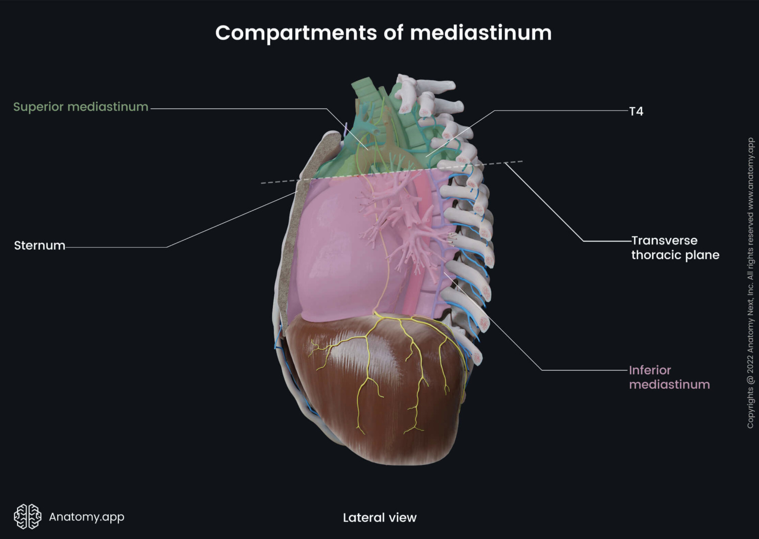
Source: Anatomy.app
What Causes Mediastinal Masses?
The most common causes of a mediastinal mass in the anterior mediastinum are thymomas, teratomas, lymphomas, and thyroid goitres. Thymomas arise from the thymus gland and can be associated with autoimmune disorders like myasthenia gravis and rheumatoid arthritis. Fibrosing mediastinitis, where fibrous tissue gradually infiltrates the mediastinum, can also occur in this region due to various causes, including infections, autoimmune disorders and previous radiation exposure.
Masses often arise in the middle mediastinum from enlarged lymph nodes, bronchogenic cysts, or vascular anomalies. These can be due to infections, inflammatory conditions, or systemic diseases like sarcoidosis.
The mediastinal masses in the posterior mediastinum are typically neurogenic tumours arising from nerve tissues. These are more common in younger people and are often benign. However, due to their proximity to the spine, they may still need to be removed.
Some risk factors, such as a family history of cancer, exposure to radiation, or underlying genetic conditions, may increase the likelihood of developing a mediastinal mass. However, in many cases, no clear risk factor is identified.
Types of Tumours
Knowing the types of tumours that can occur in the mediastinum is important in determining the treatment approach.
Thymomas are the most common tumours in the anterior mediastinum and can be benign or malignant. Lymphomas can also occur in the mediastinum and may present with systemic symptoms.
Germ cell tumours, though rare, develop from cells that produce sperm or eggs and can be found in the mediastinum. These tumours include teratomas, seminomas, and non-seminomatous germ cell tumours, each with varying prognosis.
Neurogenic tumours, which arise from nerve tissues, are often benign and located in the posterior mediastinum.
Mediastinal Tumours Symptoms
Mediastinal mass symptoms can be varied. Some people are asymptomatic, and the mass is found incidentally during an imaging scan for something else. Others may have symptoms due to the mass compressing nearby structures in the chest.
Common symptoms are chest pain, a persistent cough, shortness of breath, and/or feeling a fullness or pressure in the chest. Some patients also report hoarseness, difficulty swallowing, or swelling in the face or neck, especially if the mass compresses major blood vessels like the superior vena cava.
If the mass is malignant or linked to lymphoma, systemic symptoms such as unexplained weight loss, fever and night sweats may also be present. These symptoms need immediate evaluation.
Since many mediastinal masses do not produce symptoms in their early stages, routine imaging is important for early detection, especially in high-risk individuals.

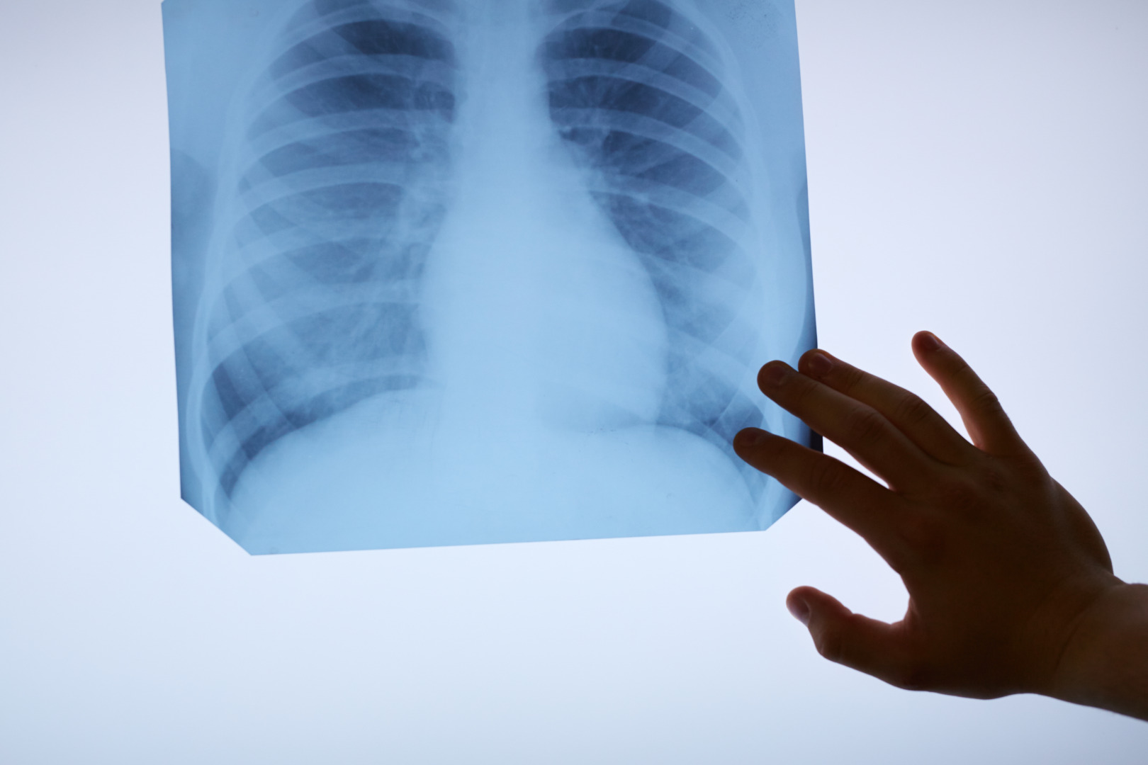
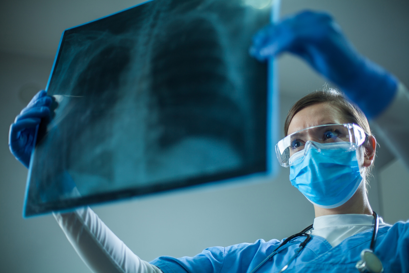
Diagnosis and Imaging
Initial evaluation usually starts with a chest X-ray, which may show a widened mediastinum or an unexpected shadow. Further imaging would be necessary for a more detailed assessment.
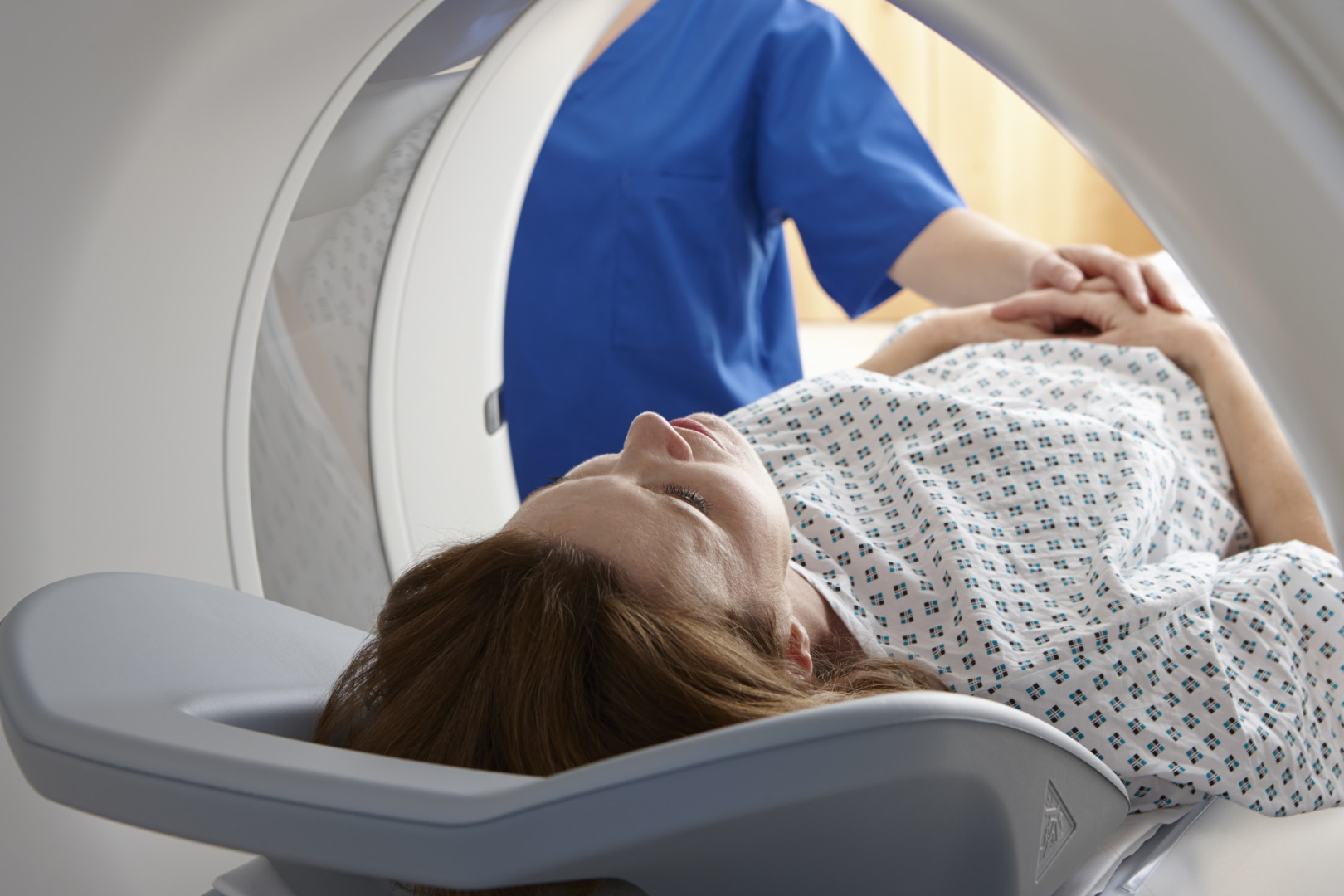
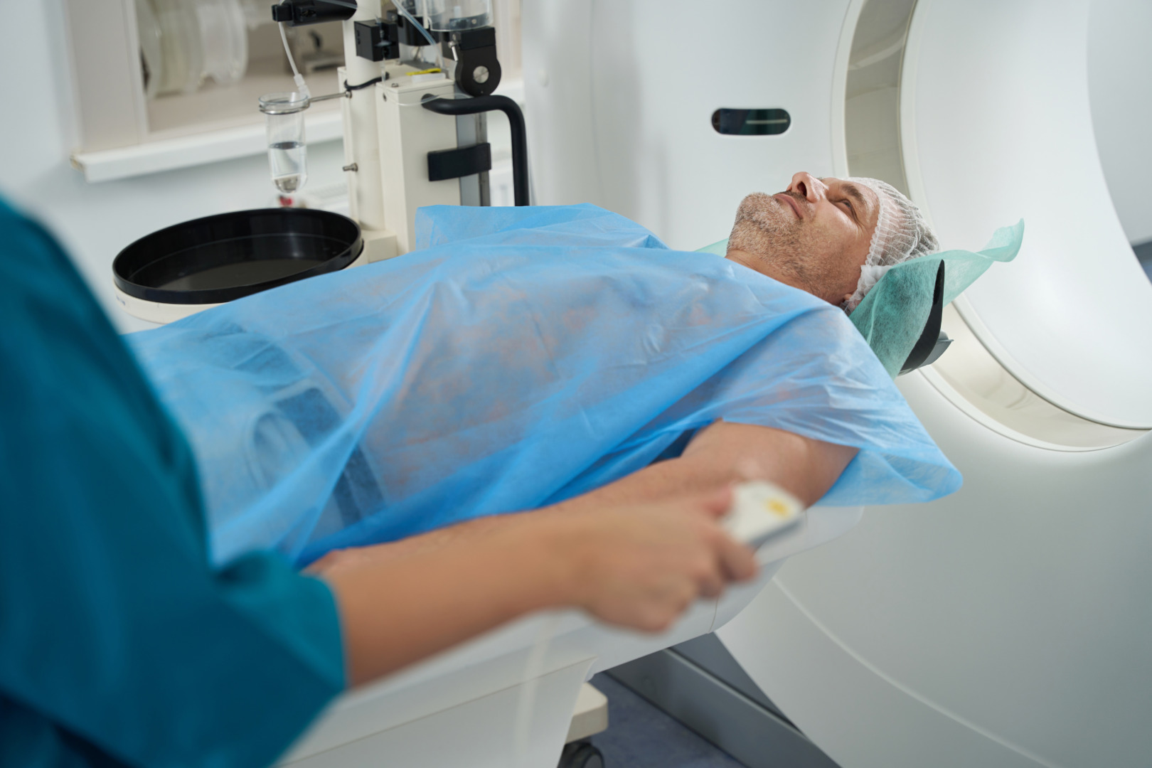
A chest CT scan is key in evaluating a mediastinal mass. It defines the size, shape and anatomical location and assesses the involvement of surrounding structures. In some cases, an MRI or PET-CT scan may be recommended to evaluate metabolic activity or further delineate soft tissue involvement.
PET is beneficial in evaluating the metabolic activity of anterior mediastinal masses, guiding biopsy or surgical planning, and differentiating between benign and malignant lesions.
Tissue sampling is often necessary to determine the nature of the mass. Depending on the location and accessibility of the mass, this can be done via a CT-guided biopsy, endobronchial ultrasound (EBUS), or mediastinoscopy. Getting adequate tissue samples during biopsies is important for accurate analysis and to avoid complications from small, fragmented specimens.
Clinical Efficacy of RATS for Mediastinal Masses
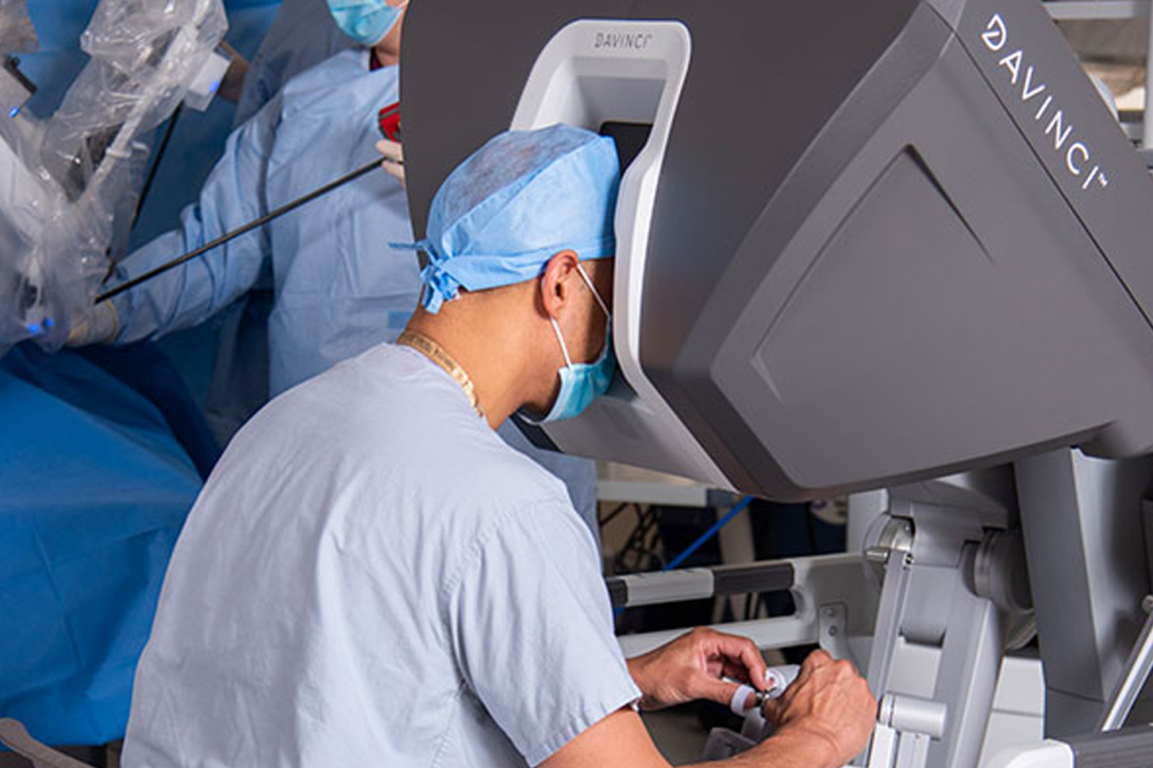
When a mediastinal mass requires surgical removal—either for diagnosis, symptom relief or curative treatment—Robotic Assisted Thoracoscopic Surgery (RATS) is a highly effective and minimally invasive option.
RATS provides surgeons with more dexterity, 3D vision and fine instrument control. These are particularly useful in the mediastinum, where delicate structures like nerves, blood vessels and the heart are packed tightly.
With RATS, thoracic surgeons can remove complex mediastinal tumours through small incisions without the need for open thoracotomy. Patients benefit from less post-operative pain, a shorter hospital stay, and faster recovery time while maintaining excellent oncological outcomes.
VATS for Mediastinal Tumour
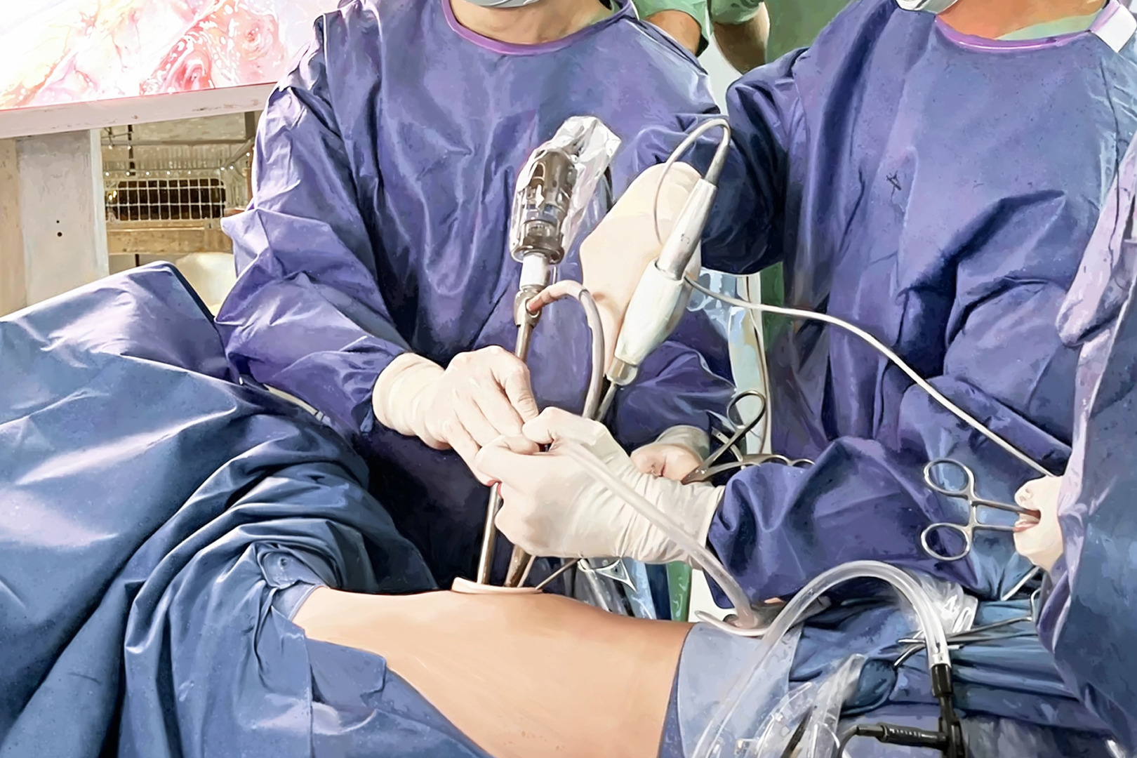
Another option for mediastinal mass treatment is VATS. Like RATS, VATS is a minimally invasive approach using a small camera and special instruments inserted through keyhole incisions in the chest.
VATS allows surgeons to visualise and remove mediastinal tumours precisely, often in cases where the mass is small, well-defined, and not attached to surrounding tissues. Depending on the type and location of the tumour and the patient’s overall health, chemotherapy and radiation may also be used as part of a comprehensive treatment plan.
Patients undergoing VATS have minimal scarring, faster return to normal activities and low complication rates. It is also ideal for patients who cannot tolerate more extensive surgery due to age or comorbidities.
In expert hands, both VATS and RATS offer beneficial outcomes, and the choice of procedure is often based on the patient’s condition, tumour location and surgical complexity.
Prognosis
The prognosis for mediastinal tumours varies widely depending on the type of tumour, its location, and the stage at which it is diagnosed. Thymomas have a good prognosis. Encapsulated thymomas have a 5-year survival rate of 96-100%. Metastatic thymomas have a poorer prognosis with 5-year survival rates of 11-50%. Germ cell tumours have variable prognosis. Teratomas have the best outcome, while seminomas and non-seminomatous germ cell tumours have a poorer prognosis.
In all cases, early detection and treatment influence the overall prognosis for mediastinal tumours.

Complications
Mediastinal tumours can cause serious complications due to their proximity to vital structures. One major complication is compression of adjacent structures like the trachea, bronchi, oesophagus, and major blood vessels like the superior vena cava (SVC). Compression of the SVC can cause SVC Syndrome, a life-threatening condition characterised by swelling and reduced blood flow. Other complications include spinal cord compression, especially with neurogenic tumours, and the spread of malignant tumours to different parts of the body. Early detection and treatment are key to preventing these complications and improving patient outcomes.

Preventing and Managing Mediastinal Mass Recurrence
Although many mediastinal masses are benign and curable with surgery, recurrence can occur, especially in cases of malignancy or incomplete resection. Therefore, long-term follow-up with imaging is important.
Patients who have undergone successful treatment may be advised to have regular CT scans and clinical reviews to monitor for any signs of recurrence. Monitoring tumour size during follow-up is essential to assess the recurrence risk and determine the treatment plan. In some cases, adjuvant therapy, such as radiation or chemotherapy, may be required to reduce the risk of regrowth, especially with aggressive tumours like lymphoma or thymic carcinoma.
Recurrence highlights the importance of choosing a highly experienced surgical team. Precision during the initial operation affects long-term outcomes.
Choosing Expert Thoracic Care
Management of mediastinal masses requires a multidisciplinary approach involving thoracic surgeons, radiologists, oncologists, radiation oncologists, and respiratory physicians. Given the complexity of the mediastinal anatomy and the range of possible diagnoses, accurate assessment and timely intervention are critical.
At Neumark Lung & Chest Surgery Centre, patients receive comprehensive care from a team of experts who specialise in minimally invasive thoracic surgery, advanced imaging and personalised treatment planning. Whether you’ve been recently diagnosed or are seeking a second opinion, our approach prioritises clinical excellence and patient well-being.
We understand a mediastinal mass diagnosis can be overwhelming. That’s why we empower patients with knowledge, support and access to the latest surgical techniques.
If you’ve been diagnosed with a mediastinal mass, are experiencing symptoms or seeking expert evaluation, contact Neumark Lung & Chest Surgery Centre in Singapore. Our experienced lung specialist and thoracic surgeon provide world-class care focusing on advanced diagnosis, minimally invasive surgery, and patient-centred care for patients with mediastinal and respiratory conditions.