

Introduction to Open Pneumothorax
Open pneumothorax, also known as a “sucking chest wound”, is a type of collapsed lung, a life-threatening condition that occurs when a wound in the chest wall allows air to pass freely between the external environment and the pleural cavity. This direct communication disrupts the normal pressure dynamics needed for lung expansion, often resulting in a partial or total lung collapse.
The severity of an open pneumothorax is determined by the size of the chest wall opening and the amount of lung compromised. If left untreated, it can rapidly progress into a tension pneumothorax where the trapped air accumulates and pushes on the lungs and heart, leading to respiratory failure and shock. Whether it be an open or tension pneumothorax, treatment from an experienced professional is necessary for the best outcomes.

Causes and Risk Factors
The most common cause of open pneumothorax is blunt or penetrating trauma to the chest. Injuries from accidents, falls, or violent incidents such as knife wounds or other chest wounds can create an opening that disrupts the seal of the chest wall. Rib fractures can also pierce the lung or pleura and allow air to enter the thoracic cavity.
In some cases, open pneumothorax can be caused by surgical procedures like chest tube placement or thoracic intervention, especially if the chest wall is weakened or damaged. Patients with chronic lung disease, such as cystic fibrosis or connective tissue disorders, may be more prone to develop this condition, especially after minor trauma. Occupational hazards, such as exposure to heavy machinery or industrial accidents, can also increase the risk of developing an open pneumothorax.

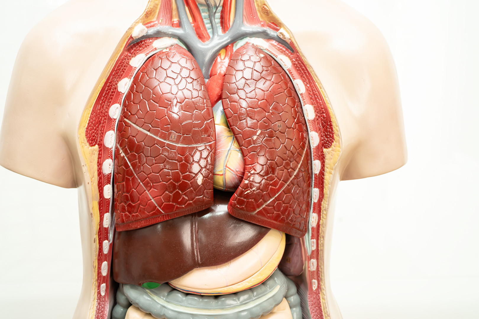
Chest Wall Pathophysiology
In a normal person, the pleural cavity maintains a negative pressure that keeps the lungs expanded during inhalation. In an open pneumothorax, this seal is lost due to the chest wall defect. So, with each breath, air enters through the external wound instead of the airway, preventing the lung from inflating properly.
In some cases, the chest wound may act as a one-way valve, known as the valve effect. Air enters during inhalation but cannot escape during exhalation. This can result in a tension pneumothorax, where pressure builds up in the chest cavity, shifting the mediastinum and compressing the heart and the opposite lung. The distinction between tension pneumothorax vs open pneumothorax lies in this trapped air effect, which can be fatal if not decompressed urgently.
Without rapid treatment, the lung will collapse and internal structures will shift, including the trachea, and the patient may develop severe hypoxia, low blood pressure, and cardiac arrest.
Clinical Presentation and Symptoms
The symptoms of open pneumothorax usually present suddenly and are often dramatic. Patients may report sharp chest pain and severe shortness of breath. A visible chest wound may be present along with a sucking or hissing sound as air moves in and out with each breath.
Breath sounds are often absent or reduced on the affected side. The chest wall may move asymmetrically, and the patient may appear pale, sweaty and anxious. In severe cases, signs of shock may develop, including tachycardia, hypotension and confusion. If respiratory failure sets in, cyanosis or altered mental status may follow.
Early recognition of these signs is crucial, especially in trauma settings where multiple injuries may mask the severity of the chest injury.

Diagnosis of Open Pneumothorax
Open pneumothorax is a clinical diagnosis made based on visible chest trauma and respiratory symptoms. In emergencies, treatment may start before imaging is done.
A physical examination will reveal a chest wound, decreased or absent breath sounds and possibly subcutaneous emphysema. A specialist may also listen for a sucking sound to help confirm air movement through the wound.
Once the patient is stabilised, a chest X-ray or CT scan can provide more detail. Imaging will confirm lung collapse, visualise the amount of air in the pleural space and assess any underlying injuries to the lungs or surrounding structures. Specifically, a chest radiograph is essential for confirming the diagnosis of open pneumothorax, evaluating its size and guiding management decisions.

Emergency and Definitive Treatment of Open Pneumothorax
Immediate open pneumothorax treatment involves closing the chest wound to prevent further air entry. A temporary dressing, often taped on three sides, allows air to escape while preventing it from re-entering. This converts the open wound into a more controlled situation, reducing the risk of a full-blown tension pneumothorax.
A chest tube is typically inserted on the affected side to evacuate air from the pleural space and allow the lung to re-expand. Oxygen therapy or mechanical ventilation may be necessary depending on the patient’s respiratory status.
In more severe cases or when the wound is large or accompanied by internal injuries, surgery is required. Complex surgical repairs, such as those for lung injuries and thoracic defects, are performed in the operating room. Surgical intervention may involve restoring the chest wall, controlling bleeding, or removing damaged lung tissue to achieve optimal outcomes.
Minimally invasive techniques, such as Uniportal Video-Assisted Thoracoscopic Surgery (U-VATS), are increasingly used in selected patients for the treatment of pneumothorax. U-VATS is performed through a single small incision, allowing for the localisation and repair of air leaks, resection of damaged lung areas, or pleurodesis to prevent recurrence. Although open traumatic pneumothorax usually requires more urgent open surgery, U-VATS may be suitable for patients with persistent air leaks or after initial stabilisation.
The benefits of U-VATS include less postoperative pain, reduced scarring, a shorter hospital stay, and a quicker return to normal activity. At Neumark, our thoracic surgery team assesses each case to determine if a minimally invasive approach is possible.

Complications and Open Pneumothorax Management
Open pneumothorax can lead to serious complications if not treated promptly. The most feared is the transition to a tension pneumothorax, which can rapidly impair cardiac function and cause death.
Other risks include infection at the wound site, persistent air leaks, respiratory failure and the development of fibrous adhesions within the pleural cavity. For patients requiring prolonged ventilation or who have large air leaks, close monitoring in the intensive care unit is often necessary.

Recovery and Long-Term Management
Recovery from an open pneumothorax depends on the size of the chest wall defect, the degree of lung injury, and whether the patient has underlying lung disease. After initial stabilisation, often involving a tube thoracostomy to remove air from the pleural space and surgical repair of the chest wall, patients need to be closely monitored to ensure the lung re-expands fully and the chest wall heals properly.
Ongoing management may involve respiratory therapy to address chest pain, improve lung function and reduce shortness of breath. For patients with chronic lung conditions such as cystic fibrosis or collagen vascular disease, long-term follow-up is essential to prevent recurrence and manage any complications.
With comprehensive management and attention to lung health, most patients can return to their normal activities. However, it is crucial to remain vigilant for any new symptoms, make necessary lifestyle adjustments, and work closely with healthcare providers to maintain optimal respiratory function.

Prevention of Open Pneumothorax
While not all cases can be prevented, there are ways to reduce the risk of open pneumothorax. Wearing seatbelts and other protective gear during high-risk activities can prevent traumatic injuries. Individuals with lung conditions should be extra cautious about activities that can cause chest trauma.

Contact Neumark for An Assessment Today
Open pneumothorax is a serious condition that requires rapid assessment and treatment. If untreated, it can become a life-threatening emergency. With proper medical care, including surgical management when needed, patients can recover fully and regain normal lung function.
At Neumark Lung & Chest Surgery Centre, our thoracic surgeons have expertise in managing complex chest injuries, including open pneumothorax. We offer emergency and elective surgery, including minimally invasive techniques, to get the best results.
If you or someone you know has had chest trauma or symptoms of lung injury, don’t wait. Contact Neumark today to schedule a consultation and begin receiving the proper care.