

What is Spontaneous Pneumothorax?
Spontaneous pneumothorax is a collapsed lung that occurs without any chest injury or medical procedure. Air escapes into the pleural space, the thin space between the lung and chest wall and the air presses on the lung, preventing it from fully expanding, which affects the breathing.
A spontaneous pneumothorax may affect part or all of one lung. The condition can start suddenly without any external cause, sometimes even while you are resting. You may develop a sudden chest pain and shortness of breath without an obvious trigger. There are two main types:
Primary spontaneous pneumothorax
Primary spontaneous pneumothorax, sometimes called idiopathic spontaneous pneumothorax, occurs in people with no known underlying lung disease. It is more common in tall, thin young adults because of their anatomy.
Secondary spontaneous pneumothorax
Secondary spontaneous pneumothorax occurs in people who already have lung disease. Chronic obstructive pulmonary disease (COPD), cystic fibrosis and idiopathic pulmonary fibrosis are among the possible causes. Secondary spontaneous pneumothorax may cause more severe symptoms because the affected person has less lung function in reserve than others.
These forms of pneumothorax differ from a traumatic pneumothorax, which follows a thoracic trauma such as a blunt injury, stab wound or other penetrating trauma. A pneumothorax caused by a medical procedure is called iatrogenic pneumothorax. A tension pneumothorax can develop when pressure builds in the pleural space and affects breathing or circulation. A tension pneumothorax is a medical emergency that needs immediate medical treatment.
Risk factors for primary spontaneous pneumothorax include smoking, a previous episode, and family history. Other risk factors include connective tissue disorders such as Marfan syndrome. A previous episode also increases the risk of a recurrent pneumothorax.

Symptoms of Spontaneous Pneumothorax
Spontaneous pneumothorax symptoms usually start suddenly. Symptoms vary depending on the size of the collapse, any existing lung disease, and your general health. Common symptoms include:
- Sudden chest pain. The pain is usually sharp, affects one side of the chest and may worsen when you breathe deeply or cough.
- Shortness of breath. Acute dyspnoea can be mild or severe. Secondary spontaneous pneumothorax may cause greater breathing difficulty in those with chronic obstructive pulmonary disease or another lung disease.
- Persistent dry cough. Some people develop a dry cough that does not settle.
A small spontaneous pneumothorax may cause few signs and can be missed. A larger pneumothorax may cause a rapid heart rate, low oxygen levels, or reduced breath sounds. Absent breath sounds, respiratory distress, bluish lips, fainting, or confusion suggest severe symptoms or clinical instability.
Go to an emergency department if your chest pain is severe, breathing becomes increasingly difficult, or symptoms worsen quickly. Call emergency services if you have signs of respiratory failure or a tension pneumothorax. Do not wait for a clinic appointment, as these symptoms are disconcerting and dangerous.
Symptoms alone cannot confirm a spontaneous pneumothorax. A chest X-ray is commonly used to look for air in the pleural space. In most cases, chest radiography confirms the diagnosis and shows how far the lung has moved away from the chest wall. If the chest radiograph is unclear but the suspicion remains high, your doctor may recommend a CT scan. The differential diagnosis includes other urgent causes of chest pain and shortness of breath.
Treatment and Management of Spontaneous Pneumothorax
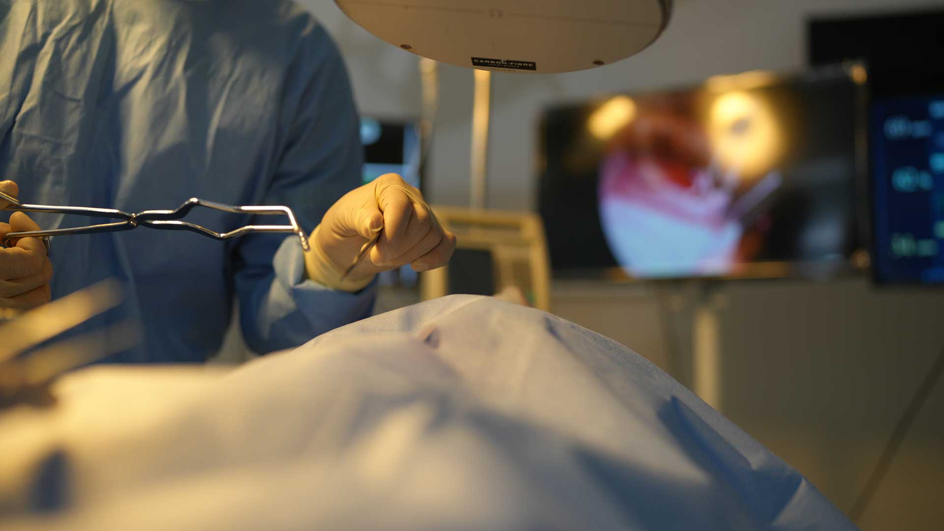
The management of spontaneous pneumothorax depends on your symptoms, stability, type, imaging findings, underlying lung disease, and whether this has happened before. Size is considered, but it is not the only factor in treating pneumothorax. The decision between conservative and interventional treatment for spontaneous pneumothorax depends on expert advice from chest physicians and thoracic surgeons, using their observations, needle aspiration, a chest tube, or surgical treatment.
Stable patients with a small primary spontaneous pneumothorax and minimal symptoms may be monitored with follow-ups and repeated chest radiography. This conservative management allows the pleural air to be absorbed while the leak closes. Use supplemental oxygen when blood oxygen is low or for other clinical reasons. This is not required for every stable patient.
Needle aspiration removes air from the pleural space through a needle or small catheter. After needle aspiration, a chest X-ray usually checks lung expansion. If needle aspiration is unsuccessful or the pneumothorax enlarges, a chest tube may be needed. A chest tube is a flexible tube placed through the chest wall into the pleural space.
A chest tube insertion allows air to leave the pleural cavity so the lung can re-expand. Chest tube placement may be used when needle aspiration is unsuitable or unsuccessful, when there is a persistent air leak, or when secondary spontaneous pneumothorax causes significant symptoms. Chest tube drainage is also more likely to be utilised when there is clinical instability. The chest tube remains in place until the air leakage has stopped and chest radiography shows an adequate lung expansion.
Chest tube complications may include pain, bleeding, infection, subcutaneous emphysema and, rarely, re-expansion pulmonary oedema. Your care team monitors the chest tube and your breathing.
For a larger collapse or recurrent spontaneous pneumothorax, your care team decides management based on your individual condition. Your team may discuss surgery after repeated episodes, when both lungs are affected, when a persistent air leak continues, or when the lung does not re-expand. Some people may prefer to avoid surgical treatment after discussing its benefits and risks.
U-VATS Surgery for Spontaneous Pneumothorax
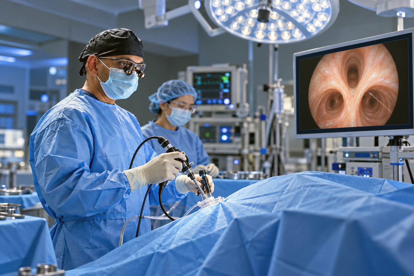
Uniportal Video-Assisted Thoracic Surgery (U-VATS) is minimally invasive surgery performed through one small incision. Video-assisted thoracoscopic surgery uses a camera and specialised instruments between the ribs. Compared with open thoracic surgery, it usually leads to less postoperative pain, a smaller scar and a shorter recovery, though results can vary.
During U-VATS for spontaneous pneumothorax, the surgeon identifies areas of air leakage. The surgeon may seal or remove blebs or bullae in the lung parenchyma to stop air from entering the pleural space. The operation may also include mechanical or chemical pleurodesis, which creates pleural adhesions between the two pleural surfaces to help prevent a recurrence.
Surgical intervention is often considered for recurrent pneumothorax or persistent air leaks. Without surgical intervention, recurrence has been reported as high as 50 percent in certain cases. Video-assisted thoracoscopic surgery (VATS) can reduce the likelihood of another episode, but no procedure removes the risk completely. The appropriate thoracic surgery depends on your age, health, lung disease, lifestyle and previous spontaneous pneumothorax treatment.
Primary spontaneous pneumothorax treatment depends heavily on the individual. Some stable patients recover with observation or needle aspiration. Others need chest tube placement. Surgery may be considered when recurrence prevention is especially important. Secondary spontaneous pneumothorax often needs closer monitoring because underlying lung disease can make breathing problems more serious.
Understand Your Options
If you have had a spontaneous pneumothorax, attend follow-up appointments and repeat imaging as advised. Smoking cessation addresses one of these risk factors. If you smoke and would like to stop, ask your care team for smoking cessation support. They can offer you advice or direct you to specialised help.
Stay active as advised during your recovery. Do not fly with an untreated spontaneous pneumothorax, as changes in air pressure can affect it. After the pneumothorax has resolved, ask your doctor when it is safe to fly. Scuba diving is generally discouraged unless a specialist confirms that a definitive procedure has addressed the risk.
Neumark Lung & Chest Surgery Centre assesses spontaneous pneumothorax, persistent air leaks and recurrent lung collapses. Management may involve monitoring, drainage, or thoracic surgery. Your care plan depends on your symptoms, imaging, lung disease and previous episodes.
Never ignore persistent chest pain or shortness of breath. Seek urgent assessment at an emergency department if symptoms return or worsen.
Frequently Asked Questions
Can a spontaneous pneumothorax heal without surgery?
Yes. Some clinically stable adults with a minimally symptomatic primary spontaneous pneumothorax can be managed with observation and follow-up imaging. Medical assessment is still required because similar symptoms can have other serious causes, and some pneumothoraces need drainage.
What is the difference between primary and secondary spontaneous pneumothorax?
Primary spontaneous pneumothorax occurs without a known underlying lung disease. Secondary spontaneous pneumothorax occurs in a person with lung disease and may cause more severe breathing problems because lung reserve is already reduced.
When is surgery considered?
Surgery may be discussed after a recurrent pneumothorax, with a persistent air leak, when the lung does not re-expand, when both sides are affected, or when recurrence prevention is especially important. The timing of surgery for spontaneous pneumothorax is individual and should be decided with a thoracic specialist.
Can spontaneous pneumothorax happen again?
Yes. Recurrence is possible, especially after a previous episode. Smoking cessation and appropriate follow-up are important. Read more about recurrent pneumothorax.