
What Is a VATS Procedure for Pleural Effusion?
A Video-Assisted Thoracoscopic Surgery (VATS) procedure for pleural effusion is a minimally invasive chest operation used to drain fluid, inspect the pleura and treat the cause when simpler tests are not enough.
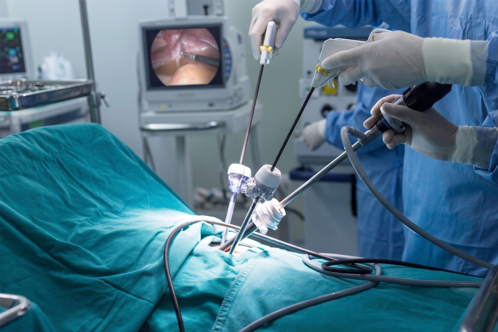
VATS is also called keyhole chest surgery. During the procedure, a thoracic surgeon makes small cuts in the chest and inserts a thin camera called a thoracoscope. The camera shows the inside of the thorax on a screen. Fine surgical instruments are then used to drain the effusion, take tissue samples and perform procedures where needed.
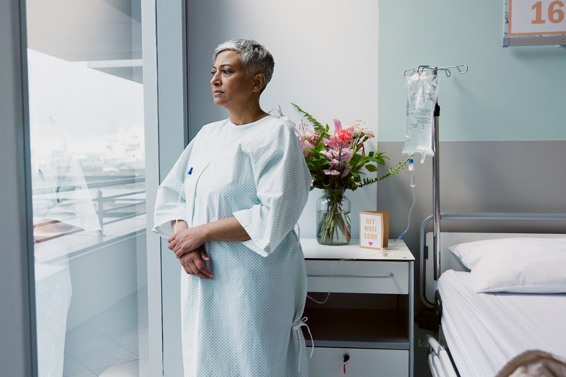
Pleural effusion means fluid has collected in the pleural space, the thin gap between the lung and the chest wall. A small amount is normal. Excess fluid can press on the lung, making it harder to breathe.
Not every effusion needs VATS. Many can be managed with medications, needle drainage, or a chest tube.
Why Might VATS Be Needed?
VATS is usually considered when the effusion keeps returning, is difficult to drain, may be caused by cancer or infection, or when doctors need a clearer diagnosis.
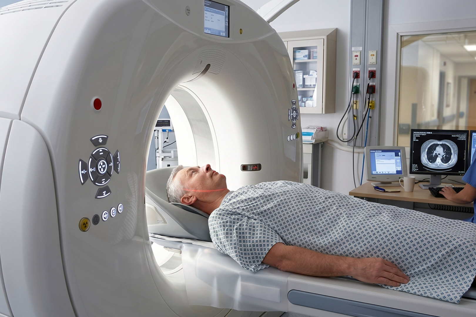
A chest X-ray, ultrasound or CT scan can show the effusion around the lung, but imaging does not always explain why it is there. A needle drainage procedure, called a thoracentesis, can remove a sample for testing. Sometimes this gives a clear answer. In other cases, the test does not confirm the diagnosis, especially when lung cancer, tuberculosis or other pleural disease is still suspected.
Video-Assisted Thoracoscopic Surgery (VATS) allows the surgeon to directly visualise the pleura, the lining around the lungs and chest wall. This can be useful when there is pleural thickening, pleural nodules, loculated pleural effusion fluid, trapped lung, or recurrent effusion buildup. By allowing a pleural biopsy under direct vision, VATS can provide an accurate diagnosis and help doctors identify cancer or infection.
VATS can be used to:
- Drain the effusion from the chest
- Take pleural biopsies
- Break down pockets of fluid or pus
- Perform talc pleurodesis
- Assess whether it can fully re-expand
This makes VATS both a diagnostic and treatment procedure.
How to Prepare for VATS
Preparation for Video-Assisted Thoracoscopic Surgery focuses on confirming the diagnosis, assessing the patient’s fitness for anaesthesia, and planning the safest and most minimally invasive approach.
Before surgery, your care team may arrange blood tests, chest imaging, heart tests and lung function tests. These help assess your overall health, your suitability for general anaesthesia, and whether VATS is the right option for you. Your doctor will also review your medications. Blood thinners may need to be stopped or adjusted before surgery.
You will usually be asked not to eat or drink for several hours before the operation. You will also have a chance to speak with the surgeon and anaesthetist. This is the right time to ask about the reason for the surgery, the expected benefits, possible risks and what recovery and follow-up appointments may involve.
What Happens During the Procedure?
During a Video-Assisted Thoracoscopic Surgery, you are asleep under general anaesthesia while the surgeon drains the effusion and treats the pleural space through small chest incisions.
You are usually positioned on your side. One lung may be temporarily deflated so the surgeon can see and work safely inside the chest. The surgeon then makes small incisions between the ribs. A camera is inserted through one incision, and surgical instruments through the others. A newer approach, uniportal-VATS (U-VATS), uses a single incision and may suit selected patients.
The surgeon may drain the effusion, inspect the lung and pleura, take biopsies and perform a talc pleurodesis. If there is an infection, thick pus or pockets of fluid, the surgeon may clear these areas and wash out the chest cavity. If there is a trapped lung, the surgeon may remove thickened lung tissue from the surface so the entire lung can re-expand in selected cases.
At the end of the operation, one or more chest drains are usually placed. These drains remove air, fluid or blood while the lung re-expands.
Benefits and Limitations
Compared with open surgery, VATS can provide both diagnosis and treatment in a single procedure, with smaller incisions.
The main benefit is that the surgeon can directly visualise the pleura and perform targeted pleural biopsies. This can improve diagnostic accuracy and yield a more precise diagnosis when fluid testing alone has not provided a clear answer. VATS can also treat the effusion at the same time by draining the effusion and performing pleurodesis where appropriate.
As VATS uses smaller cuts than a thoracotomy, recovery often involves less pain and is faster than open surgery. VATS allows for faster recovery, with many patients recovering sooner, walking earlier, and returning to normal activities sooner. Compared with a thoracotomy, VATS generally causes fewer complications.
VATS still has limits. It may not be suitable if the patient is too unwell for general anaesthesia, if the pleural disease is very advanced, or if the lungs cannot re-expand. In some cases, the surgeon may need to perform additional surgery or convert to open surgery if that is safer.
Risks and Recovery
Recovery after VATS depends on the cause of the effusion, the procedure performed and how well the lung re-expands.
Most patients stay in the hospital for a few days after VATS, although this varies. The chest drain may stay in place for 24 to 48 hours or longer, depending on how much fluid or air is draining. Chest X-rays are usually used to check that the lung has expanded before the drain is removed.
Pain is usually managed with regular pain relief. Walking, deep breathing and coughing exercises are important because they help reduce the risk of pneumonia, blood clots and pulmonary embolism. Patients may feel tired for a few weeks after surgery.
Possible risks and complications include bleeding, air leak from the lung (a pneumothorax), chest or wound infection, pain, blood clots, pneumonia and recurrence of the effusion.

How Neumark Can Help
Neumark Lung & Chest Surgery Centre can help determine whether VATS is the right procedure for an effusion and provide minimally invasive surgery when it is appropriate.
Neumark specialises in pleural and thoracic surgery with a multidisciplinary approach led by Dr Harish Mithiran, senior consultant thoracic surgeon at Gleneagles and Mount Alvernia hospitals. For patients with an effusion, this means a careful review of scans, symptoms, fluid test results and overall health before recommending surgery.
If you have a recurrent effusion, unexplained fluid, a suspected malignant pleural effusion, or an infection that is not settling, contact Neumark for a consultation.