
What is a Loculated Pleural Effusion?
A loculated pleural effusion is fluid around the lung that becomes trapped in separate pockets rather than flowing freely.
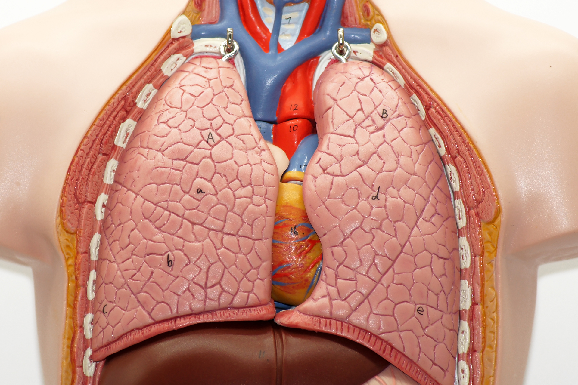
The pleura is the thin lining around the lung and the inside of the chest wall. A small amount of fluid normally helps these layers move smoothly during breathing. A pleural effusion happens when too much fluid collects in this space. In a simple effusion, the fluid is usually free-flowing. In a loculated pleural effusion, the fluid is divided into pockets by strands of tissue called septations or adhesions.
Loculated effusions occur when inflammation causes scar tissue to form. Adhesions then create walls that trap fluid in compartments. These pockets can make the effusion harder to drain. A needle or chest tube may remove fluid from one pocket but leave other pockets behind.
Loculation can develop when fluid stays around the lung for some time or when the pleura (the lining around the lung) becomes irritated or inflamed. As the area tries to heal, scar-like tissue can form, dividing the fluid into separate pockets.
This can happen with pneumonia, pleural infection, tuberculosis, bleeding into the chest, cancer, previous chest or abdominal surgery, or with long-term inflammation. Some conditions, such as a complicated pneumonia-related effusion, malignant pleural effusion, recurrent cancer-related fluid, or chronic fluid from cirrhosis, can make pocketing more likely.
Malignant pleural mesothelioma can also cause scarring and thickening of the pleura, which may trap fluid and make drainage more difficult.
Repeated fluid build-up can irritate the pleural surfaces. Over time, this irritation can lead to inflammation and scar-like tissue. The fluid may then become trapped in separate pockets rather than moving freely.
Common causes include congestive heart failure, pneumonia, tuberculosis, and cancer. Lymphoma and other blood-related cancers can also cause pocketed fluid around the lung.
Less common causes include nephrotic syndrome, rheumatoid arthritis, viral infection, blunt chest trauma, penetrating chest injury, and complications after trauma. Conditions such as heart failure, cirrhosis, and other chronic illnesses often cause free-flowing fluid, but the fluid can become loculated if an infection or ongoing inflammation develops.
Treating loculated effusions is more complex than treating free-flowing effusions because the fluid does not occupy a single open space.
Symptoms of a Loculated Pleural Effusion
Symptoms of loculated pleural effusion depend on the amount of fluid, the cause, and whether the lung can expand. Symptoms of a loculated pleural effusion vary widely.
Some people have mild symptoms, especially if the pockets are small. Others feel short of breath when fluid builds up in the lungs.
Common symptoms include:
- Breathlessness
- Dyspnea
- Shortness of breath
- Chest heaviness
- Pleuritic chest pain along the lateral chest wall
- Dry cough
- Fever
- Tiredness
Dyspnea, the medical term for shortness of breath or difficulty breathing, is among the earliest signs in many patients and can worsen as fluid accumulates. Patients describe dyspnea on exertion, at rest or with a sudden onset.
Fatigue can result from reduced oxygen intake. When the lung cannot expand fully, breathing takes more effort, and normal activity feels more tiring.
Symptoms may also give clues about the cause a loculated pleural effusion:
- Fever, chills, and feeling unwell may suggest infection or empyema (pus in the pleural space).
- Weight loss, poor appetite, and recurrent fluid may raise concern for cancer.
- Night sweats and a chronic cough may suggest tuberculosis.
- Sudden chest pain and breathlessness may point to a pulmonary embolism.
A loculated effusion can feel more persistent than a simple one. Symptoms may not improve after one drainage if fluid remains trapped in other pockets.

When to See a Thoracic Surgeon
You should see a thoracic surgeon when pleural fluid is loculated, recurrent, infected, difficult to drain, or may be suspicious for cancer.
A loculated pleural effusion should not be managed as though it were a simple fluid collection. Thoracic specialist review is important because treatment may need targeted drainage, intrapleural medicine, biopsy, thoracoscopy, or surgery.
A thoracic surgeon is important if drains for a loculated pleural effusion or chest tube insertion are not working, if the time interval since chest tube insertion is long, or if the infection is becoming organised. Surgical review for a clearer diagnosis is also important when the diagnosis is unclear, and tissue is needed to rule out cancer, tuberculosis, latent tuberculosis, or other diseases.
You should seek urgent medical care if you have severe breathlessness, high fever, worsening chest pain, confusion, blue lips, or rapidly worsening symptoms. These may indicate serious infection, complications, or a large effusion.

Diagnosis of a Loculated Pleural Effusion
Diagnosis relies on imaging, fluid analysis, and sometimes direct inspection of the pleural space.
Medical history guides the workup. This covers chest trauma, pneumonia, heart failure, cancer and prior procedures. A careful medical history helps narrow down the most common causes.
A chest X-ray may show fluid around the lung, but it may not clearly show whether the fluid is trapped in pockets. This is why loculated effusions can be missed or underestimated on a standard X-ray.
Ultrasounds are often more helpful because they can show the separate pockets and thin internal strands, called septations. Ultrasounds also help doctors find the best place to insert a needle or drain (usually along the side of the chest) and are especially useful because fluid moves with gravity, so the imaging can show where it has settled.
A computed tomography (CT) scan gives a more detailed view of the chest. It can show trapped fluid, pleural thickening, lung collapse, infection, a lung mass, scar tissue along the pleural surfaces, or a trapped lung. If there is blood in the pleural space, CT scans may also show signs of a clot.
Loculated fluid is quite common in more complex pleural effusions. One CT scan estimate found loculation in 58% of exudative pleural effusions. This matters because exudative effusions are often associated with infection, inflammation, cancer, or other active disease processes. When fluid is loculated, it may need closer assessment because simple drainage may not remove all the trapped pockets.
A thoracentesis may be done to take a fluid sample for testing or to remove fluid and ease breathlessness. The fluid is checked for signs of infection, cancer cells, protein, sugar, acidity, cell count, and inflammation. These results help doctors determine whether the fluid is due to an infection, cancer, heart failure, or another condition. In loculated pleural effusions, thoracentesis may not drain much fluid if the needle reaches only one pocket or if the fluid is thick. This is why ultrasound or CT guidance is important.
If the cause remains unclear, a pleural biopsy may be recommended. This can be done with imaging guidance or during thoracoscopy, where a camera is placed into the chest so the pleura can be seen directly.

Non-Surgical Management of a Loculated Pleural Effusion
Non-surgical treatment focuses on draining the right pockets, treating the underlying disease, and helping the lung re-expand. Interventional pulmonology offers image-guided drainage that may help many patients avoid surgery.
Treatment depends on the underlying condition. If the cause is pneumonia or community-acquired pneumonia, antibiotics are essential. If the fluid is large, infected, or causing shortness of breath, drainage is usually needed.
Sometimes a chest tube alone is not enough because the fluid is thick or septated. Medicine may be placed through the tube to help break down the pockets and improve drainage. This may include fibrinolytic therapy in selected infections. Fibrinolytic therapy helps dissolve scar-like strands and thin the fluid.
For malignant pleural loculated effusions, treatment depends on whether the lung can expand, how fast the fluid returns, and the patient’s overall condition. Options include repeated drainage, an indwelling pleural catheter, cancer treatment, or pleurodesis if the lung expands well. In patients with non-expandable lungs or loculated malignant effusions, an indwelling pleural catheter may be more effective than pleurodesis.
Treatment of loculated pleural effusion should be individualised to the underlying disease. The aim is not just to remove fluid once but to prevent fluid recurrence while treating the cause. It is to improve breathing, control infection if present, clarify the diagnosis and reach an accurate diagnosis, and reduce the chance of repeated procedures.
Surgical Options for a Loculated Pleural Effusion
Surgery is considered when a loculated pleural effusion does not drain well, causes a trapped lung, remains infected, or requires a tissue diagnosis.
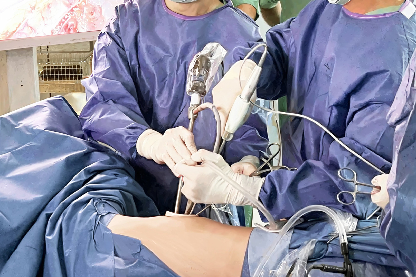
Video-Assisted Thoracoscopic Surgery (VATS) is often the preferred surgical option. It uses small incisions and a camera to look inside the chest. Through VATS, the surgeon can drain fluid pockets, break adhesions, remove infected material, take pleural biopsies, wash out the pleural space, and perform pleurodesis when appropriate.
VATS is especially useful for empyema and complex parapneumonic effusions when antibiotics and chest tube drainage are insufficient. It can also help when a malignancy is suspected, but fluid testing has not given a clear answer. Direct visualisation of the pleura allows for a targeted biopsy and treatment in the same setting.
A medical thoracoscopy may also help in selected cases. It allows a direct visualisation of the pleura and can be used for adhesiolysis, biopsy, drainage and talc pleurodesis. The choice between a medical thoracoscopy and VATS depends on the patient’s condition, the complexity of the fluid, the need for general anaesthesia, and the likelihood of requiring more extensive surgical treatment.
A thoracotomy is an open chest operation. It is used less often today but still has a role when the disease is advanced, the fibrous peel is thick, the lung is trapped, or minimally invasive surgery cannot safely clear the problem. In these cases, decortication may be needed to remove thick fibrous tissue from the lung surface so that the lung can re-expand.
Risks and Recovery
Recovery after treatment depends on the cause, the thickness of the fluid, the procedure used, and how well the lung re-expands.
After a thoracentesis, many patients recover quickly, but loculated effusions may not drain completely with a single procedure. Possible complications include pain, bleeding, infection, pneumothorax (or pneumothorax-related complications), and incomplete drainage.
After a chest tube drainage, patients may stay in the hospital while fluid drains and imaging is repeated. Intrapleural medicines can improve drainage in selected cases but may cause pain, bleeding, or fever. The care team monitors the response and decides if further treatment is needed.
After a thoracoscopy or VATS, recovery is usually faster than an open surgery. Many patients stay in the hospital for one to three days after a thoracoscopy, depending on the cause, drainage needs, and how well the lung re-expands. Patients are encouraged to walk early, do breathing exercises, and use pain medicine so they can cough and breathe deeply. A chest drain often stays in place for a short time.
A thoracotomy requires a longer recovery because the incision is larger and the operation is more extensive. It is reserved for more advanced or complex diseases.
How Neumark Can Help
Neumark Lung & Chest Surgery Centre helps by identifying the cause of a loculated pleural effusion and selecting the appropriate treatment, ranging from targeted drainage to minimally invasive surgery.
Neumark specialises in pleural and thoracic conditions with a multidisciplinary approach led by Dr Harish Mithiran, senior consultant thoracic surgeon at Gleneagles and Mount Alvernia hospitals. For patients with loculated pleural effusion, this means carefully reviewing ultrasound and CT findings, fluid results, symptoms, and overall health before recommending treatment.
Some patients need antibiotics and image-guided drainage. Others need intrapleural medicine, a biopsy, VATS, pleurodesis, or decortication. When surgery is appropriate, Dr Harish may use minimally invasive thoracic techniques to improve drainage, obtain a diagnosis, and help the lung re-expand where possible.
If you have been told you have a loculated pleural effusion, or if fluid around the lung keeps coming back or does not drain properly, contact Neumark for a consultation.