
What Is Robotic-Assisted Thoracic Surgery Thymectomy?
Robotic-Assisted Thoracic Surgery (RATS) thymectomy is a minimally invasive operation to remove the thymus gland using robotic instruments controlled by a thoracic surgeon.
The goal of the surgery depends on the reason for the operation. For a thymoma or another thymic mass, the aim is a complete resection of the thymus and tumour, often with the surrounding fatty tissue. In myasthenia gravis, the aim is an extended thymectomy, which removes the thymus and nearby mediastinal fat that may contain thymic tissue. Either way, an experienced professional will walk you through the process and what to expect from your RATS thymectomy.
In adults, a thymectomy is most often considered for thymoma, other selected thymic malignancies, and myasthenia gravis. Traditionally, this operation was performed through the conventional trans-sternal approach with a sternotomy. RATS offers a different route. Instead of one large incision, the surgeon works through small ports using a camera, fine surgical instruments and robotic arms.
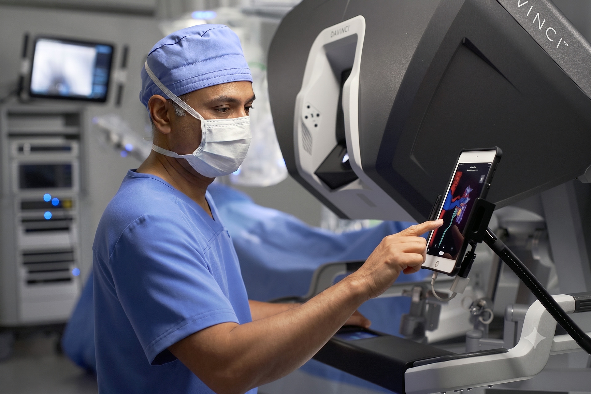
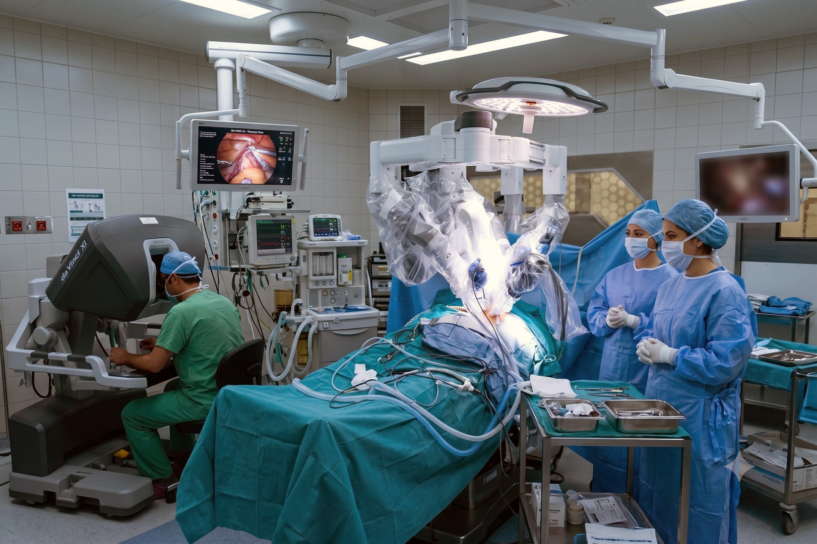
The robotic system does not operate on its own. The surgeon remains in control throughout the procedure at the robotic console. With the da Vinci surgical system, the surgeon uses a magnified, three-dimensional view and wristed instruments that move with high precision in narrow spaces. This is especially useful in the anterior mediastinum, where the thymus gland lies close to the heart, lungs, phrenic nerve, major blood vessels and the innominate vein.
Symptoms Requiring Thymectomy
The symptoms linked to a robotic-assisted thymectomy usually come from the underlying thymic condition, not from the thymus itself being removed.
Many thymomas are found incidentally on a scan and cause no symptoms. When symptoms do occur, they may include chest discomfort, coughing, shortness of breath, or a feeling of chest pressure. Larger tumours can sometimes press on nearby structures and cause more obvious symptoms.
Others are referred for surgery because of myasthenia gravis, a chronic autoimmune disorder that causes muscle weakness that tends to worsen with activity and improve with rest. Common symptoms include drooping eyelids, double vision, difficulty swallowing, slurred speech, weak arms or legs, and tiredness. In more serious cases, breathing muscles can be affected by myasthenia gravis.
When to See a Thoracic Surgeon
You should see a thoracic surgeon when a thymic tumour is suspected, when thymectomy is being considered for myasthenia gravis, or when imaging shows an anterior mediastinal mass.
A thoracic surgeon helps answer the practical questions that matter most. Is the mass likely to be resectable? Is minimally invasive surgery suitable? Does the patient need a biopsy first, or is direct surgical treatment more appropriate? How extensive should the resection be? A thoracic surgeon helps assess whether RATS is appropriate or whether video-assisted thoracoscopic surgery (VATS) or if open surgery would be safer. All of these answers vary from patient to patient.
Diagnosis
Diagnosis before a RATS thymectomy focuses on identifying the reason for surgery, defining the anatomy, and deciding whether a minimally invasive approach is suitable.
Most patients begin with imaging, such as computed tomography (CT). A contrast-enhanced chest CT scan shows the thymus, the size of any anterior mediastinal mass, its relationship to nearby vessels, including the innominate vein, and whether the lesion appears confined or invasive. Not every case is suitable for RATS. Very large tumours, obvious invasion into surrounding structures, or situations where major vascular reconstruction may be needed, can make open surgery the better surgical approach.
Robotic-Assisted Thoracic Surgery Thymectomy Using the da Vinci System
The da Vinci surgical system is a critical component of RATS for thymectomies, as it provides enhanced visualisation and precise instrument control for optimal results.
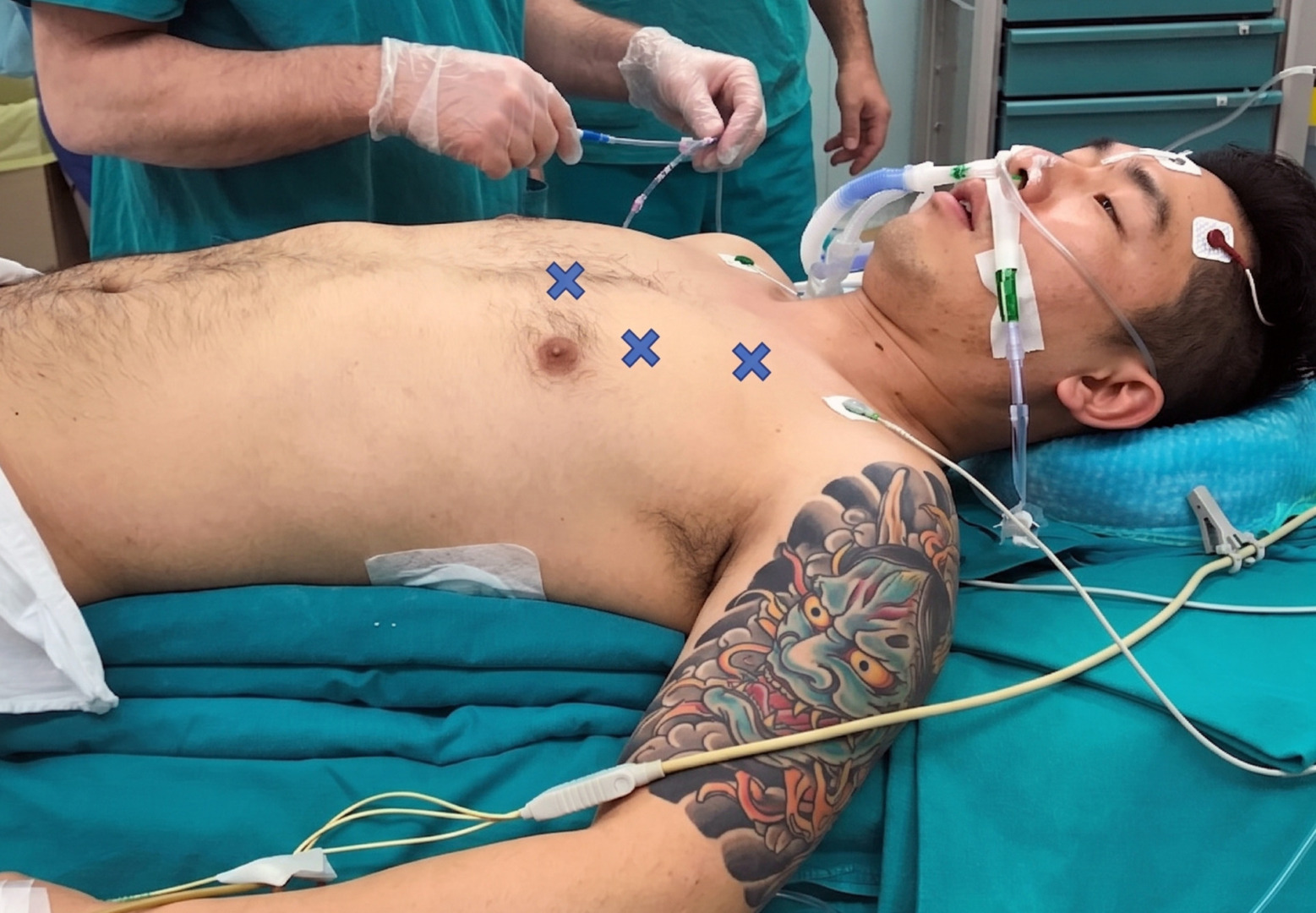
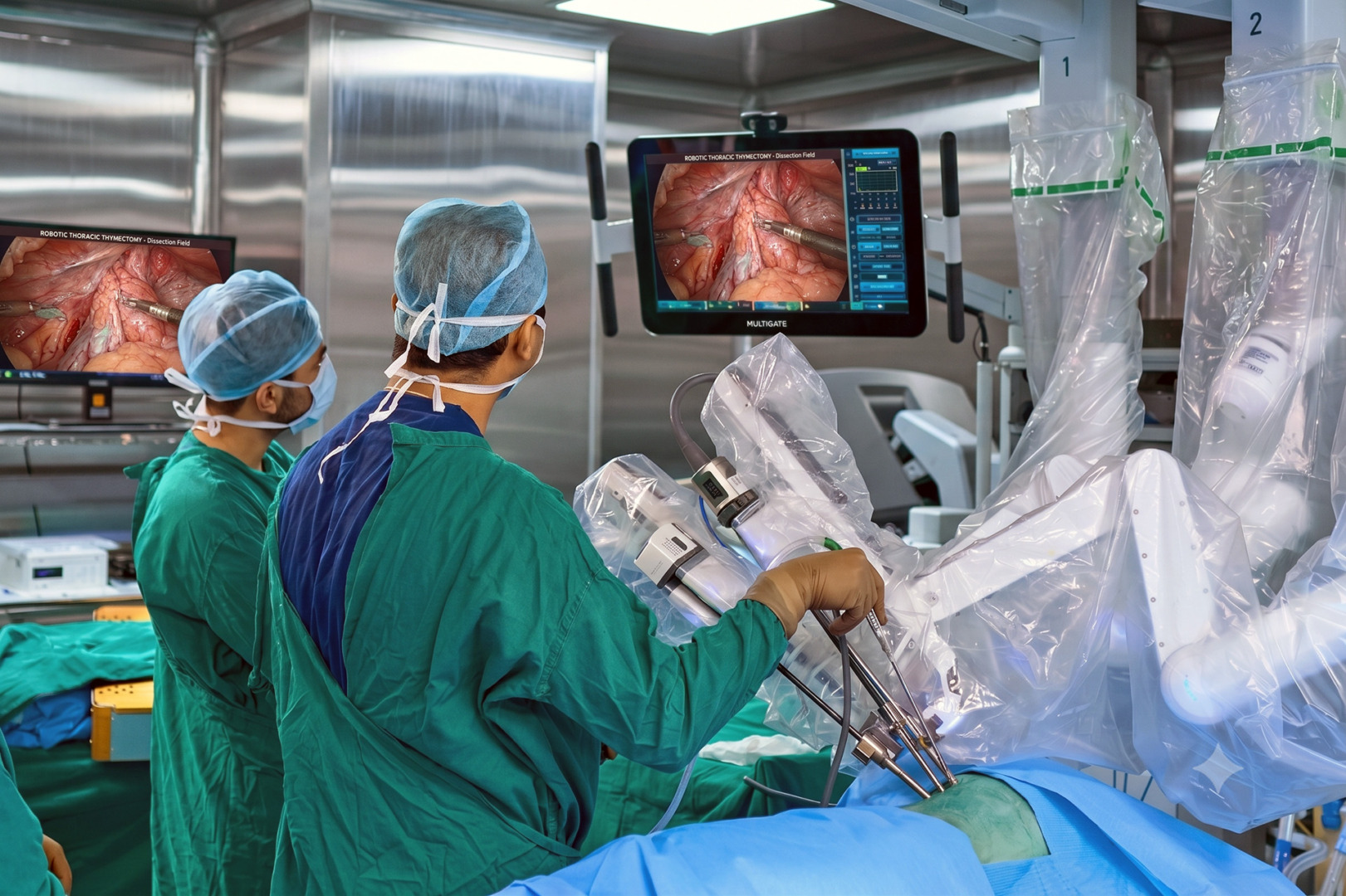
After general anaesthesia, the patient is positioned on the operating table, with the patient’s position optimised to allow better access to the anterior mediastinum. Ports are placed along the anterior axillary line, and the robotic arms are introduced through these ports. The da Vinci robotic system provides a stable three-dimensional view of the surgical field, and the robotic arms manoeuvre surgical instruments with wristed precision in the narrow chest.
The operation involves careful dissection around the thymus gland, both thymic horns, and the surrounding fatty tissue. Protection of the right and left phrenic nerves is particularly important because a phrenic nerve injury can affect diaphragmatic movement and breathing.
For myasthenia gravis, the operation is usually planned as an extended thymectomy. For thymoma, including Stage I and II thymoma, the goal is a complete resection without rupturing the tumour capsule. If there is any concern during the operation that a robot-assisted approach may compromise safety, conversion to an open procedure may be necessary. Your specialist will speak to you about this possibility.
Risks and Recovery
A robot-assisted thymectomy is generally well tolerated in suitable patients, but it is still a major thoracic surgery procedure and carries real risks.
Risks include bleeding, infection, an air leak, postoperative pain, phrenic nerve injury, injury to blood vessels, injury to the lung, and anaesthetic complications. Postoperative complications in myasthenia gravis include perioperative respiratory weakness, which is why coordination with a neurologist and anaesthesiologist is essential.
Most patients spend a short time in the hospital, and postoperative pain is usually less than with an open surgery. A chest tube is placed during thoracoscopic surgery, and the chest tube is removed when drainage is minimal and the lung is well expanded. A second chest tube may occasionally be needed if an air leak persists. Many people are up and walking early and return to light activity sooner than after a sternotomy.
Recovery also depends on why the surgery was done. In thymoma, follow-up focuses on pathology, margin status, staging, and surveillance imaging. Thymectomy for myasthenia gravis does not produce an immediate remission for all patients, and improvement is often gradual. Careful follow-up remains important.
How Neumark Can Help
Neumark Lung & Chest Surgery Centre helps by assessing whether a Robot-Assisted Thoracic Surgery (RATS) thymectomy is the right operation and by planning treatment around the patient, not just the scan.
Neumark specialises in minimally invasive thoracic surgery with a multidisciplinary approach led by Dr Harish Mithiran, senior consultant thoracic surgeon at Gleneagles and Mount Alvernia hospitals.
For patients with thymoma, an anterior mediastinal mass, or myasthenia gravis requiring surgical treatment, this means a careful imaging review, a thoughtful assessment of surgical suitability, and a clear discussion of whether RATS, VATS, or open thoracic surgery would provide the safest and most effective treatment.
In thymectomy for myasthenia gravis and thymic malignancies, the focus is on a complete resection, phrenic nerve protection, and a minimally invasive recovery whenever the surgical approach allows. The best operation is the one that is safest, oncologically sound, and suited to the individual patient. There is no one-size-fits-all approach, which is why the expertise and experience of a trained professional like Dr Harish Mithiran are key to performing a thymectomy.
If you have been told you may need a thymectomy, or if you have myasthenia gravis or a suspected thymic tumour and want a thoracic surgical opinion, contact Neumark Lung & Chest Surgery Centre for a consultation.