

What Is Transudative Pleural Effusion?
The pleura consists of two layers: the visceral pleura, which covers the lung surface; and the parietal pleura, which lines the inside of the chest wall. A small amount of fluid normally sits in the pleural space between the pleural layers to help the lungs move smoothly during breathing. Pleural effusion happens when too much fluid collects in the pleural space.
Specifically, transudative pleural effusion occurs when fluid around the lungs is caused by changes in pressure or protein balance in the body, rather than by inflammation of the pleura itself. This is what makes pleural transudates different from exudative pleural effusions, which are more often caused by chronic inflammation, infection, rheumatoid arthritis, cancer, or direct pleural disease.
This usually means that hydrostatic pressure inside blood vessels is too high or that decreased plasma oncotic pressure due to low serum protein levels allows fluid to leak out of vessels, or both. Excess fluid then shifts into the pleural space.
Congestive heart failure raises hydrostatic pressure in pulmonary capillaries, causing fluid to seep into the pleural space. Other causes of transudative pleural effusion also include:
- Liver cirrhosis with ascites, causing hepatic hydrothorax
- Nephrotic syndrome
- Renal failure
- Pulmonary embolism
- Severe low serum protein
- Hypothyroidism
- Renal vein thrombosis
- Ovarian hyperstimulation syndrome
- Fluid movement related to peritoneal dialysis
Symptoms of transudative pleural effusion depend on how much fluid has accumulated and how quickly it develops. The distinction matters because the treatment is different. A transudative effusion is usually managed by treating the underlying heart, liver, kidney, or a systemic condition. Drainage of accumulated fluid may help relieve symptoms, but it does not address the root cause unless the underlying cause is also treated.
Symptoms
The symptoms often overlap with the underlying cause of transudative pleural effusion. A person with congestive heart failure may have bilateral pleural effusion along with swollen legs, tiredness, rapid weight gain, or breathlessness that worsens when lying down. A person with liver cirrhosis causing hepatic hydrothorax may have abdominal swelling from ascites. A person with kidney disease or nephrotic syndrome may have generalised swelling, foamy urine, or changes in urination.
Small effusions may not cause any respiratory symptoms. They may be found incidentally on a chest radiograph or computed tomography (CT) scan done for another reason. However, larger effusions can compress the lung, causing respiratory symptoms. Symptoms may include shortness of breath, chest heaviness, dry cough, reduced exercise tolerance, and difficulty lying on your back.
Chest pain is less typical in a simple transudative effusion because the pleura is not usually inflamed. Sharp pain that worsens with breathing, accompanied by fever, chills, or feeling very unwell, may suggest another cause, such as an infection, pulmonary embolism, or an exudative pleural effusion. Importantly, exudative pleural effusions from bacterial pneumonia, parapneumonic effusion, Mycobacterium tuberculosis, malignant pleural disease, or oesophageal rupture more often cause pleuritic pain and systemic illness. These symptoms need a prompt medical review.

When to See a Thoracic Surgeon
Most transudative effusions are first managed by physicians, cardiologists, respiratory specialists, kidney specialists, or liver specialists, depending on the cause, as the primary treatment is usually medical. A thoracic surgeon is not always needed.
Thoracic surgical review becomes more important when the effusion keeps returning despite adequate medical treatment, when it causes repeated hospital admissions, or when the diagnosis is not fully clear. It is also useful when imaging suggests a trapped lung, pleural thickening, loculated fluid, or another condition that does not fit a simple transudate.
Some underlying causes of transudative pleural effusion need immediate treatment. Patients should seek urgent care if breathlessness is severe, oxygen levels are low, there is sudden or intense chest pain, or there are symptoms such as fainting, confusion, blue lips, or rapidly worsening breathing. A massive pleural effusion can seriously affect breathing and require urgent assessment.
At Neumark Lung & Chest Surgery Centre in Singapore, Dr Harish Mithiran helps assess when a pleural effusion requires procedural treatment and when the safest course is to avoid surgery and focus on the underlying medical condition.

Diagnosis
Diagnosis starts with imaging and is confirmed by analysing the pleural fluid when the cause is unclear, or the symptoms are significant.
One of the first questions is whether the fluid is transudative or exudative. A transudative pleural effusion usually happens because of a body-wide fluid imbalance, such as heart failure, liver disease or kidney disease. An exudative pleural effusion is more often associated with inflammation, infection, cancer, or disease affecting the pleura itself.
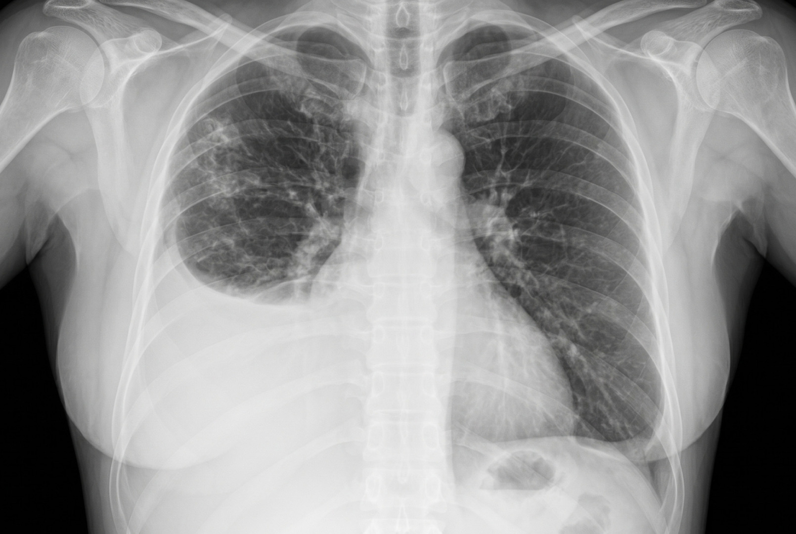
A chest X-ray can show fluid around the lung. A lateral decubitus chest X-ray will help confirm the presence of freely layering fluid and estimate its volume. A second lateral decubitus view may be obtained if the results are inconclusive. Ultrasound scans help estimate the amount of fluid and can guide safe drainage. CT scanning assesses the pleural space and may be used when the effusion is large, one-sided, recurrent, or associated with other concerning findings. These imaging tests help identify features such as pleural thickening, a trapped lung, or bilateral pleural effusion that inform the diagnosis.
Doctors use pleural fluid analysis to help distinguish between them. The test compares protein and enzyme levels in the pleural fluid with levels in the blood. These results help show whether the fluid is more likely to be a transudate or an exudate. Thoracentesis is a procedure that removes a small sample of fluid from around the lung using a needle. The sample is tested in a lab to determine why the fluid has collected.
Other tests for transudative pleural effusion may include measuring the fluid’s pH and glucose levels, cell count, and assessing for signs of infection or cancer. A low pleural fluid pH may suggest infection or a complicated pneumonia-related effusion. For some patients concurrently taking diuretics, the fluid may appear more inflammatory than it really is, so doctors may use additional blood and fluid comparisons to avoid a misclassification.
The goal is not just to name the type of effusion. It is to find the cause. Once the cause is clear, treatment can be matched to the underlying condition.
Even so, clinical judgement still matters. For example, a patient treated with diuretics for heart failure may have fluid results that look more like an exudate despite a heart-related cause. This is sometimes called a pseudo-exudate. In such cases, the doctor reviews the full picture, including symptoms, scan findings, blood tests, response to treatment, and other medical conditions.
Tests may also include kidney and liver function tests, blood protein levels, thyroid tests, cardiac markers, echocardiography, and tests for infection or cancer if the presentation is unusual.
Non-Surgical Management
Treatment of transudative pleural effusion usually focuses on addressing the underlying disease causing the fluid buildup.
In heart failure, treatment may include diuretics, fluid and salt management, and optimisation of the heart medications. As the heart condition improves, the pleural fluid may reduce. In kidney disease or a nephrotic syndrome, treatment may involve controlling the fluid overload, managing protein loss, adjusting medications, or initiating dialysis as needed.
In liver cirrhosis with hepatic hydrothorax, the treatment may include salt restriction, diuretics, ascites management and specialist liver care. Patients undergoing peritoneal dialysis may develop pleural effusion via a pleuroperitoneal communication, necessitating adjustments to peritoneal dialysis technique or volume. Pulmonary embolism causing pleural effusion is treated with anticoagulation to address the underlying pulmonary circulation problem.
Therapeutic thoracentesis can provide relief when the effusion is large enough to cause breathlessness. It removes fluid directly from the pleural space and can quickly improve breathing. However, if the underlying cause remains active, the fluid may return. That is why repeated drainage without a broader treatment plan is not ideal.
For recurrent or refractory transudative effusions, longer-term options may be considered. These can include repeated planned drainage, an indwelling pleural catheter in selected patients, or pleurodesis in carefully chosen cases. The best option depends on the cause, the expected course of illness, lung expansion, infection risk, and the patient’s overall condition.
Surgical Options
Surgery is not the usual first-line treatment for transudative pleural effusion, but Video-Assisted Thoracoscopic Surgery (VATS) or a thoracotomy may be considered in selected complex cases.
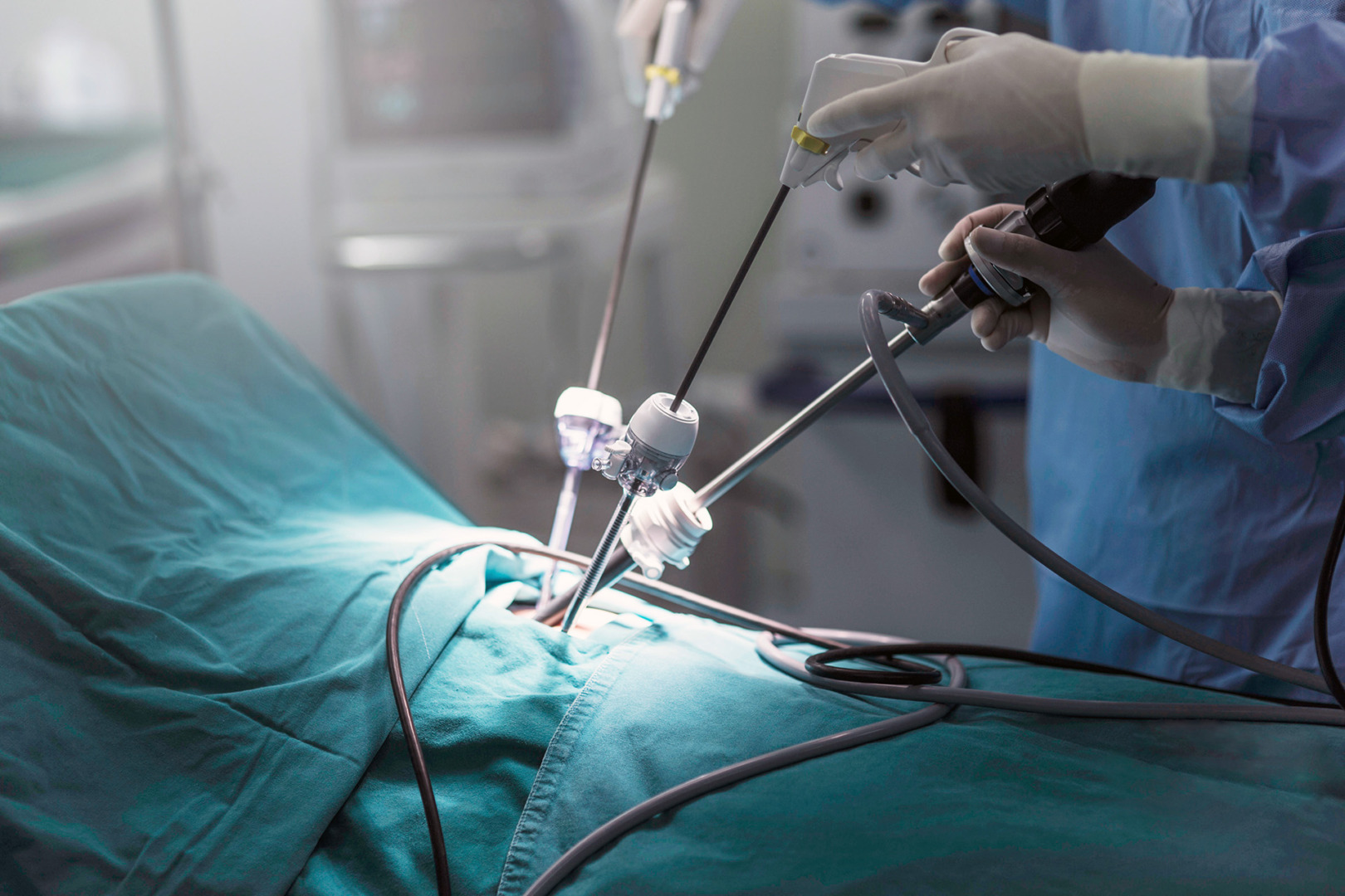
VATS allows the surgeon to inspect the pleural space through small incisions using a camera. It may be used when the diagnosis is uncertain, when the effusion behaves atypically, or when a pleural biopsy is needed to rule out malignancy or another pleural disease. VATS may also be used for pleurodesis if recurrent fluid is causing major symptoms and the lung can still expand well. In malignant pleural effusions from lung cancer or other cancers, VATS pleurodesis and chest tube drainage can help manage the pleural space. Malignant effusion from lung cancer or mesothelioma often requires an indwelling pleural catheter or pleurodesis for long-term control.
A thoracotomy is an open chest operation and, in rare cases, may be used for transudative pleural effusion. It may be considered when there is complex pleural disease, dense fibrous tissue and scarring from chronic inflammation, a trapped lung requiring decortication, or findings suggesting the effusion is not a simple transudate after all. As thoracotomy is more invasive, it is reserved for situations where less invasive options are unlikely to work or are unsafe.
Risks and Recovery
Recovery depends more on the underlying cause of the effusion than on the pleural fluid alone.
After a thoracentesis, patients are usually monitored for complications such as pneumothorax, bleeding, pain, infection, or a re-expansion of symptoms if a large volume of fluid was removed. Many patients feel relief from rapid breathing, but some require repeat imaging and follow-up.
Recovery after VATS tends to be faster than after an open surgery. Patients may need a chest drain for a short period, pain control, breathing exercises, and follow-up imaging. Recovery from a thoracotomy is longer because the incision is larger and the operation is more invasive.
The main risk of recurrence comes from the underlying condition. If heart failure, kidney disease, liver disease, hepatic hydrothorax, or low blood protein remains uncontrolled, fluid can build up again in the pleural space. Trapped lung may develop if the pleural space is repeatedly infected or if fibrous tissue accumulates. Preventing recurrence, therefore, means controlling the primary disease, monitoring symptoms, and choosing surgical pleural procedures only when they add clear benefit.
How Neumark Can Help
Neumark Lung & Chest Surgery Centre specialises in minimally invasive thoracic surgery with a multidisciplinary approach led by Dr Harish Mithiran, senior consultant thoracic surgeon at Gleneagles and Mount Alvernia hospitals in Singapore. For patients with pleural effusion, this means carefully reviewing imaging, pleural fluid analysis results, symptoms, and the likely cause before recommending a procedure.
In many cases, the right decision is not surgery. It may be better to work with the patient’s cardiologist, nephrologist, hepatologist, or respiratory physician to manage the underlying disease. In other cases, drainage, pleural biopsy, VATS pleurodesis, or another thoracic procedure may be appropriate.
If you have been told you have this condition, contact Neumark for a consultation. A timely specialist review can clarify the cause, relieve symptoms where possible, discuss whether surgery is right for you, and help reduce the risk of recurrence.