

What Is Malignant Pleural Effusion?
Malignant pleural effusion is a build-up of fluid and cancer cells in the space between the lung and chest wall.
The lungs and the inner surface of the chest wall are covered by thin linings called the pleura. The visceral pleura covers the lung, while the parietal pleura lines the chest wall. Together, they enclose the pleural cavity. A small amount of fluid normally sits between these layers to allow smooth lung movement during breathing, and lymphatic vessels drain any excess fluid. A pleural effusion occurs when too much fluid collects in the pleural space, either from increased pleural fluid production or from reduced drainage.
A pleural malignancy may develop when cancer cells spread to the pleural lining, producing pleural metastases that obstruct normal fluid drainage or trigger chronic inflammation, leading to further fluid buildup. Malignant pleural disease is usually a sign of advanced disease, but the outlook and treatment options vary from person to person. Tumour growth on the pleural surface is the underlying driver, and a malignant effusion can recur until that tumour growth is controlled.
Sometimes the first sign of cancer is not a visible tumour, but fluid that keeps returning in the chest cavity around the lung. The cancers most often linked with malignant pleural effusion include lung cancer, such as small cell lung cancer, breast cancer, lymphoma and other haematological malignancies, ovarian cancer, and mesothelioma. In many cancer patients, the effusion reflects metastatic disease from a primary tumour elsewhere, and identifying the primary malignancy guides treatment.
Malignant pleural effusion treatment usually focuses on easing breathlessness, controlling fluid buildup, improving comfort, and treating the underlying cancer where possible. The optimal plan depends on symptoms, cancer type, lung expansion, expected treatment response, patient factors, and the patient’s overall health.
Symptoms
The most common symptom of malignant pleural effusion is shortness of breath. This happens because the fluid presses on the lung, preventing it from expanding fully. Some people notice breathlessness only when walking or climbing stairs. Others feel short of breath even at rest, especially when the effusion is large.
Other symptoms may include chest heaviness, chest pain, a dry cough, tiredness, reduced appetite, and weight loss. Some patients also develop a pericardial effusion around the heart, which can add to breathlessness and chest pain.
Some patients find it harder to lie flat. Symptoms may develop gradually, but they can also worsen quickly if fluid builds up over a short time.
Symptoms are not always caused by the fluid alone. They may also come from the cancer itself, infection, anaemia, blood clots, or reduced general fitness.

When to See a Lung Specialist
You should see a lung specialist when pleural fluid is linked to cancer, keeps coming back, or causes significant breathlessness.
A specialist review is important if a scan shows a unilateral pleural effusion and there is a known cancer diagnosis. It is also important if the fluid is unexplained, bloodstained, recurrent, or associated with pleural thickening, nodularity, or nodules.
You should seek urgent medical care if your breathlessness is severe, symptoms are worsening quickly, or your chest pain is sudden or intense. A large effusion can make breathing difficult and may need prompt drainage.
A thoracic surgeon becomes involved when the diagnosis is uncertain, when a tissue biopsy is needed, when the lung does not expand after drainage, or when a longer-term fluid-control option is being considered. These options may include pleurodesis, an indwelling pleural catheter, or minimally invasive surgery.

Diagnosis
The diagnosis of malignant pleural effusion starts with medical imaging and is confirmed by testing the pleural fluid or pleural tissue.
Assessment begins with a physical examination and a review of symptoms. As breathlessness and pleural fluid may have many causes, the differential diagnosis includes heart failure, pleural infection, pulmonary embolism, and benign effusions, as well as pleural malignancy. A careful differential diagnosis guides which tests are needed, and the physical examination helps assess the size of the effusion.
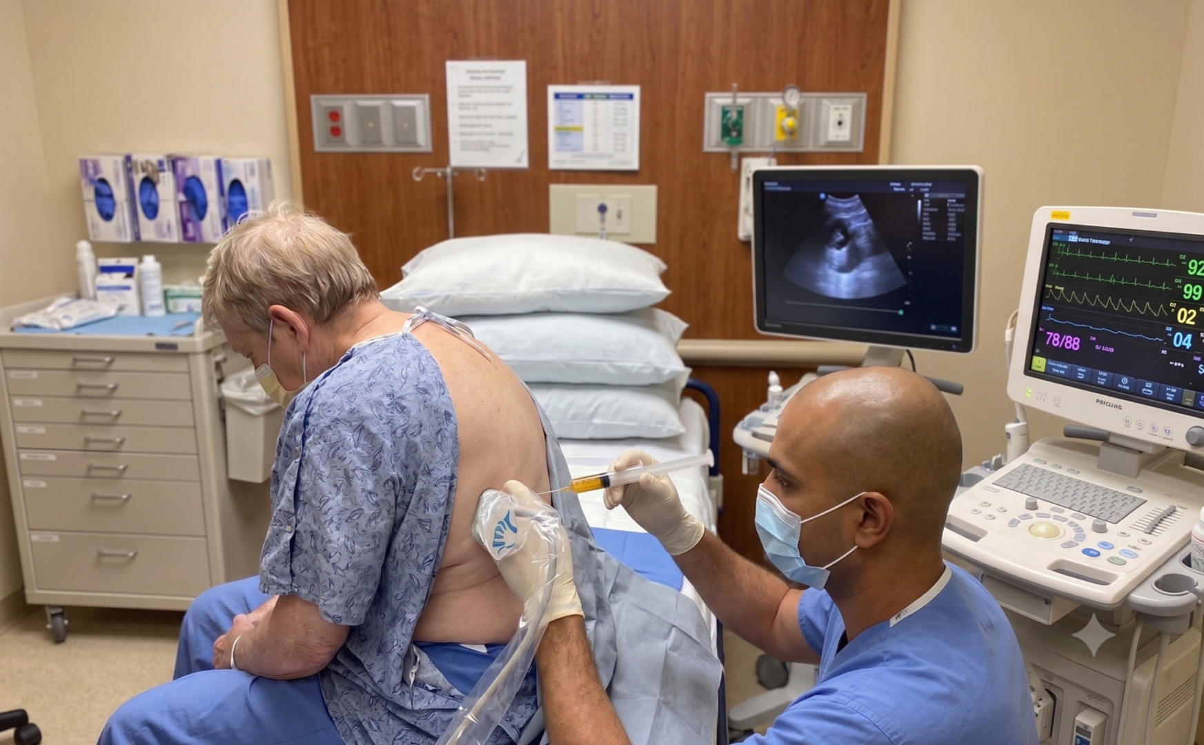
A chest X-ray may show fluid at the base of the lung, and it is often the first imaging test. A thoracic ultrasound helps confirm the amount of fluid and guides safe drainage. CT, or computed tomography, scanning gives more detail. Together, a chest X-ray and CT scans can show pleural thickening, pleural nodularity, pleural nodules, lung masses, lymph nodes, and whether the lung is trapped by tumour or scarring. A pleural thickening of more than 1 cm and nodular pleural thickening raise concern for malignancy, so this pattern of pleural thickening is taken seriously.
Thoracentesis is often the first procedure. During thoracentesis, a malignant pleural effusion assessment, a needle is used to remove fluid from around the lung. This can improve breathing and provide a sample for pleural fluid analysis. The lab checks the fluid for cancer cells, malignant cells, infections, protein levels, and cell count. Malignant effusions are usually exudative and high in protein, and pleural fluid analysis helps distinguish them from other causes.
A pleural fluid test may confirm the diagnosis, but not always. Sometimes cancer or tumour cells are not seen in the fluid even when cancer is still suspected, so a definitive diagnosis may require tissue. If the diagnosis remains unclear, a pleural biopsy may be needed to confirm pleural involvement. This can be done with image guidance or during a thoracoscopy, where a small camera is placed into the chest so the pleura can be seen directly and tissue samples can be taken.
A diagnosis also includes assessing whether the lung expands after fluid removal. This is important because some patients have a lung that cannot fully re-expand due to a tumour, scarring, or a trapped lung. Pleural manometry during fluid drainage can help detect an unexpandable lung by measuring pressure as fluid is drained.
Non-Surgical Management
Non-surgical management focuses on draining the fluid, easing breathlessness, providing symptom relief, and controlling recurrence with the least burden to the patient.
The simplest treatment is therapeutic thoracentesis. Fluid is removed with a needle to relieve breathlessness. This can work well if the effusion is the first episode, if symptoms are mild, or if doctors need to see whether drainage improves symptoms.
If the fluid returns, a repeated thoracentesis may be required. Repeated procedures also carry risks such as pain, bleeding, pleural infection, and pneumothorax, which is air leaking around the lung. For recurrent malignant pleural effusion, two common longer-term options are pleurodesis and an indwelling pleural catheter.
- Pleurodesis aims to seal the pleural space so fluid cannot collect again. This chemical pleurodesis uses a substance such as sterile talc (talc pleurodesis) placed into the pleural space, usually through a chest tube or during thoracoscopy, followed by chest tube drainage. For successful pleurodesis, the lung usually needs to re-expand after drainage, and success is lower when the lung is trapped. Pleurodesis failure is more likely with an unexpandable lung, so the risk of failure is assessed before this option is chosen.
- An indwelling pleural catheter is a soft tube that stays in the chest and allows fluid to be drained at home. Pleural catheters can be helpful for people whose fluid keeps returning, people who prefer to avoid repeated hospital visits, or those whose lungs do not fully expand. Pleural catheters give patients greater control because at-home fluid drainage reduces the need for hospital visits. In some cases, regular drainage through these pleural catheters can also lead to spontaneous pleurodesis, where the pleural space seals over time. When the lung re-expands, pleural catheters can achieve pleurodesis success without talc.
Cancer treatment also matters. Systemic treatments such as chemotherapy, immunotherapy, targeted therapies, radiotherapy, or hormonal treatment may help control fluid in some cancers. This systemic treatment may shrink tumour growth on the pleura. However, local pleural treatment is often still needed when breathlessness is significant.

Surgical Options
Surgery is considered when a malignant pleural effusion needs diagnosis, pleurodesis, fluid control, or the treatment of a trapped lung.
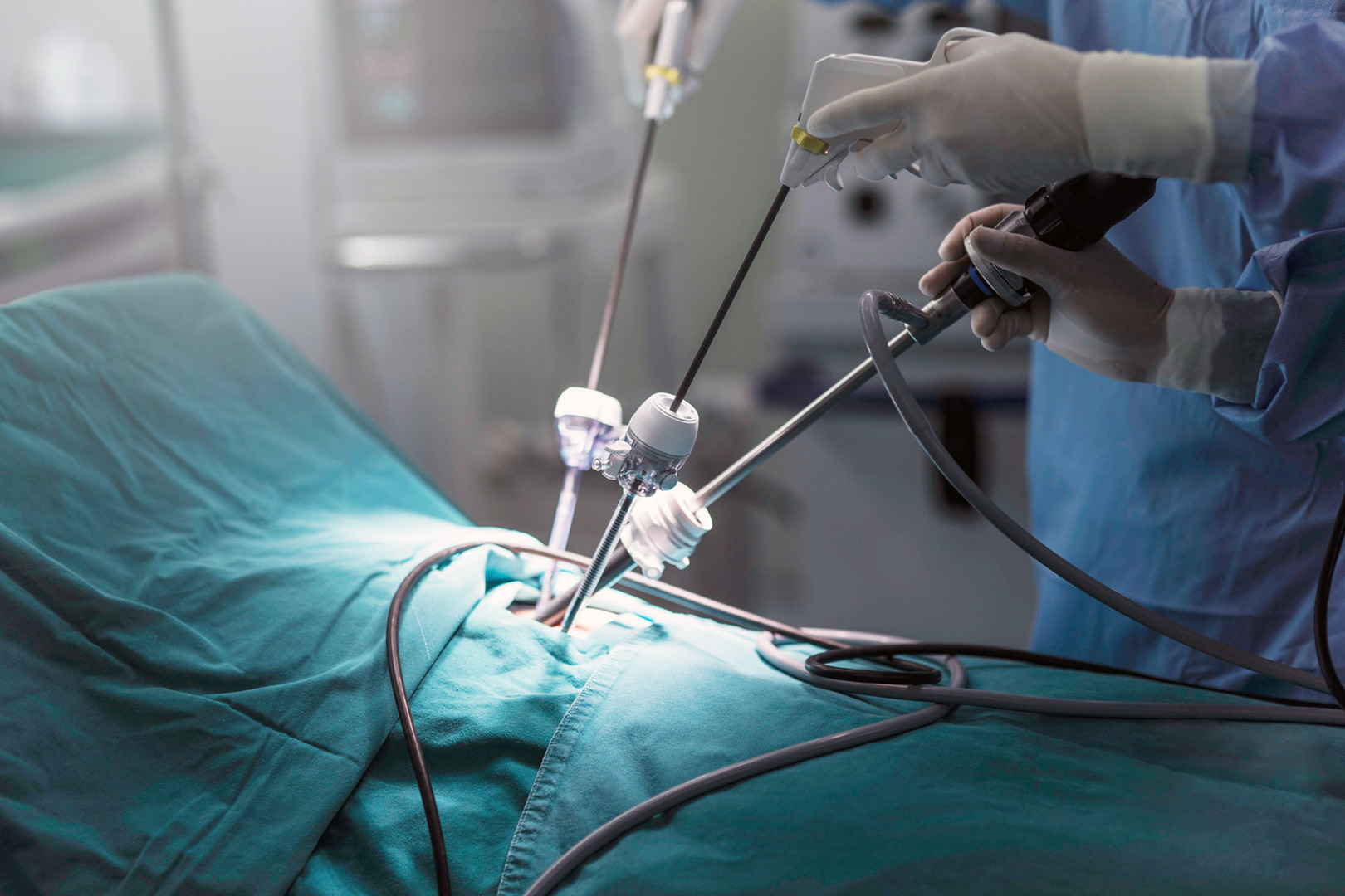
Video-Assisted Thoracoscopic Surgery, or VATS, is the main minimally invasive surgical approach. VATS uses small incisions and a camera to look inside the chest cavity. The surgeon can drain fluid, inspect the pleura, take targeted biopsies, perform talc pleurodesis, and assess lung expansion.
VATS is especially useful when pleural fluid tests have not confirmed the diagnosis, but cancer remains likely. It allows diagnosis and treatment to happen during the same procedure in selected patients.
Surgery may also be considered if the lung is trapped and symptoms are severe. In selected cases, decortication may be used to remove a thick rind from the lung surface so the lung parenchyma can expand better. This surgery is not suitable for every patient.
Open surgery through a thoracotomy is less commonly used for malignant pleural effusion alone because it is more invasive. It may be needed in selected complex cases where minimally invasive thoracic surgery is not safe or complete enough.
The aim of surgery is not always to cure. In many patients, the goal is symptom control, better breathing, a clearer diagnosis, or fewer repeat procedures.
Risks and Recovery
Recovery after malignant pleural effusion treatment depends on the procedure, the cancer, and how well the lung re-expands.
After a thoracentesis, many people feel better quickly. Some have mild soreness at the needle site. Possible risks include bleeding, pleural infection, low blood pressure, pneumothorax, or reexpansion pulmonary oedema, a form of pulmonary injury, if a large amount of fluid is drained too quickly. In rare cases, this can progress to acute respiratory distress syndrome, so reexpansion pulmonary oedema is avoided by draining in stages.
After chest tube drainage, patients may need to stay in the hospital for monitoring. Chest discomfort, fever and pain can occur after pleurodesis. A chest tube usually remains in place until fluid output decreases and the lung is fully expanded.
After VATS, recovery is usually faster than after an open surgery. Patients are encouraged to walk early and use breathing exercises. A chest tube may stay in place for a short time. Pain control is important so the patient can breathe deeply, cough, and move safely.
Malignant pleural effusion can recur, especially if the underlying cancer remains active. As it usually reflects advanced disease, it carries a poor prognosis, and median survival is measured in months for many cancer types, though this varies with the primary malignancy and treatment response.
How Neumark Can Help
Neumark Lung & Chest Surgery Centre helps patients with malignant pleural effusion by identifying the cause, relieving breathlessness, providing symptom relief, and selecting the most appropriate long-term fluid-control option.
Neumark specialises in pleural and thoracic conditions with a multidisciplinary approach led by Dr Harish Mithiran, senior consultant thoracic surgeon at Gleneagles and Mount Alvernia hospitals. For patients with malignant pleural effusion, this means careful review of scans, pleural fluid analysis, biopsy needs, lung expansion, symptoms, and overall fitness.
Some patients need thoracentesis and monitoring. Others need pleural biopsy, VATS, talc pleurodesis, indwelling pleural catheters, or surgery for a trapped lung. The right choice depends on the clinical picture and on what will best improve breathing and quality of life.
If you have been told you have a malignant pleural effusion, or if fluid around the lung keeps coming back, contact Neumark for a consultation.