What Is a Spiculated Lung Nodule?
Lung, or pulmonary, nodules are among the most common incidental findings on routine lung cancer screening CT scans.
Nodule shape is one of the features linked with a higher chance of malignancy. Ground glass nodules, ground glass opacity, part-solid nodules, solid nodules, and rounded nodules all have different risk profiles. Solid nodules with solid components may carry a higher risk. Part-solid lung nodules, including part-solid and ground-glass lung nodules, may behave differently from solid nodules. Cancerous and noncancerous lung nodules can look similar, which is why tests are important.
Lung specialists or thoracic surgeons will determine whether the detected pulmonary nodules are cancerous, noncancerous, or benign. Many lung nodules are benign; noncancerous findings include infection, inflammatory conditions such as rheumatoid arthritis, and scarring.
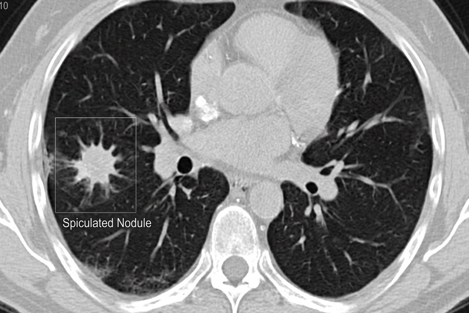
Lung nodules have varying characteristics and shapes. A spiculated lung nodule is a lung nodule with spiky or radiating edges on imaging, which raises concern for cancer but does not confirm it. A spiculated appearance is more suspicious than a smooth one, but it is not a diagnosis by itself. Some benign conditions can look spiculated, including prior infection, inflammation, scarring and fungal disease.
Your care team of physicians and your doctor will review your CT scan, imaging report and health history, including your family history and whether you are a current smoker. Imaging tests, scans and additional tests help determine the tissue’s shape and characteristics, as well as whether a tissue sample is needed. Contrast enhancement on a CT scan will also help determine whether a finding is cancerous.
If diagnosed early, cancerous lung nodules can often be treated effectively. That is why spiculated lung nodules often require further testing.
Symptoms
Spiculated lung nodule symptoms are often absent from early-stage cases because many small lung nodules are typically too small to cause any symptoms at all.
Most lung nodules are found incidentally on CT scans conducted for other reasons. When symptoms do occur, they may come from the underlying cause rather than the nodule itself. Possible symptoms include:
- Persistent cough
- Shortness of breath
- Chest pain
- Coughing up blood
- Recurrent chest infections
- Fatigue
- Unexplained weight loss.
In Singapore, this matters because lung cancer remains the third most common cancer in both men and women. Early screenings can help, especially if you are at risk.

When to See a Lung Specialist or Thoracic Surgeon
A spiculated border of a lung nodule warrants prompt specialist review, especially if the nodule is new, growing, larger, located in an upper lobe, or found in someone older, with a smoking history, prior cancer, or with concerning symptoms. A thoracic surgeon may become involved early when a nodule is difficult to biopsy, when the risk of cancer is high enough that surgery may provide both diagnosis and treatment, or when a patient wants a definitive opinion on the safest next step.
Diagnosis
Diagnosing a spiculated lung nodule means estimating the likelihood of cancer and deciding whether surveillance, a PET-CT scan, biopsy or surgery is the right next step.
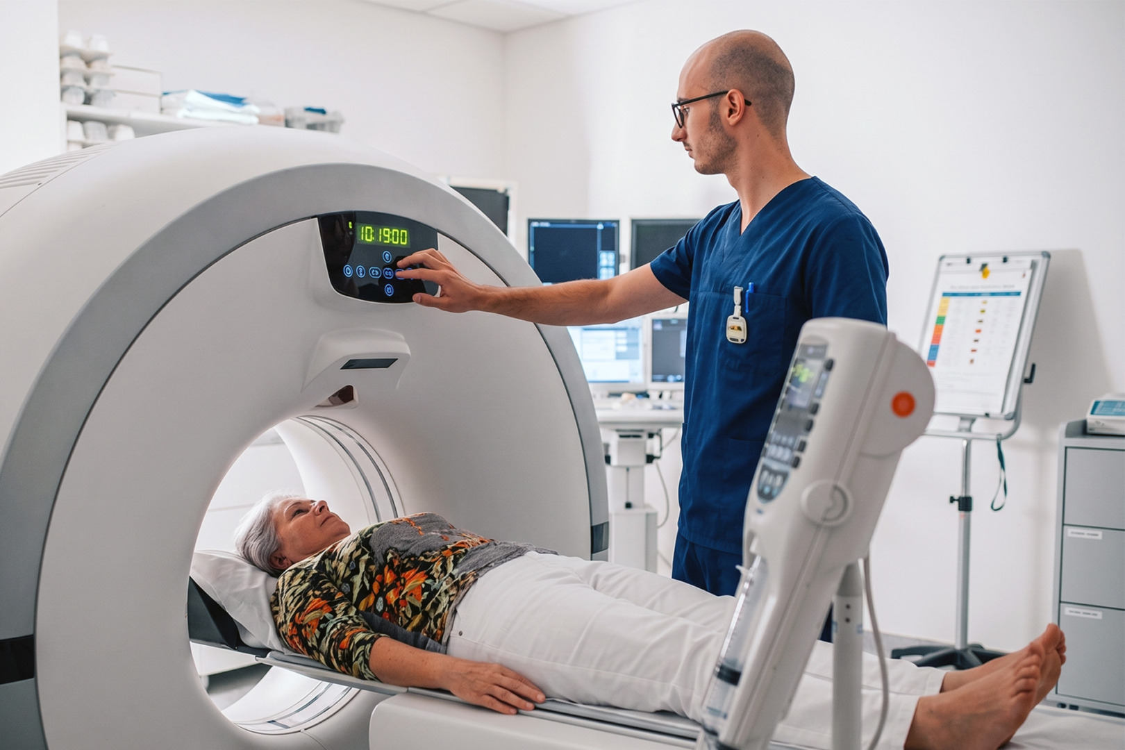
The first step is a careful review of the chest CT scan. The specialist examines the nodule’s exact size, density, location, growth pattern and margin. A comparison with previous scans is crucial because stability over time is reassuring, while growth over time increases concern. Solid nodules that remain stable for at least two years are less likely to be malignant, although subsolid nodules can change more slowly and may need longer observation.
Doctors also weigh personal risk factors. Validated prediction models use variables such as the age, smoking history, previous cancer incidence, nodule size, and the location to estimate the probability of malignancy of the lung nodule.
If the nodule is concerning enough, further tests may include a PET-CT scan, bronchoscopy, or a CT-guided needle biopsy. PET-CT scans can help assess the metabolic activity in selected lung nodules, though they are less useful for very small nodules, small lesions, or some slow-growing tumours. A bronchoscopy is often more useful for lung nodules closer to the airways, while a CT-guided biopsy is often chosen for lung nodules nearer the outer lung. If these tests are unlikely to give a reliable answer, surgery may be the best diagnostic option.
Non-Surgical Management
Many spiculated nodules can be managed without surgery when the overall risk is low enough and follow-up is reliable.
For some patients, the safest plan is active surveillance with repeat CT scans at set intervals. The goal is to see whether the nodule stays stable, resolves, or changes in a way that makes the cancer more or less likely. This avoids overtreatment of benign nodules while still identifying malignancy early, when a cure is most likely.
Non-surgical care may also include treatment for an infection or inflammatory process if the clinical picture supports it, along with smoking cessation and the management of any other lung disease. This is one reason the question “Is a spiculated nodule always cancer?” should be answered carefully. The answer is no, but that does not mean it is not cancer either. Every case is different. A spiculated nodule raises suspicion, but decisions should be based on the nodule’s full radiological and clinical context.
Surgical Options
Surgery is usually considered when the nodule is highly suspicious, enlarging, technically difficult to biopsy, or already proven to be cancerous.
The operation depends on the size and location of the lesion and on how much lung can be safely preserved. A wedge resection may be used to remove and diagnose a small peripheral nodule. A segmentectomy or lobectomy may be more appropriate when cancer is strongly suspected or confirmed. Lymph node sampling may also be needed to guide staging and further treatment.
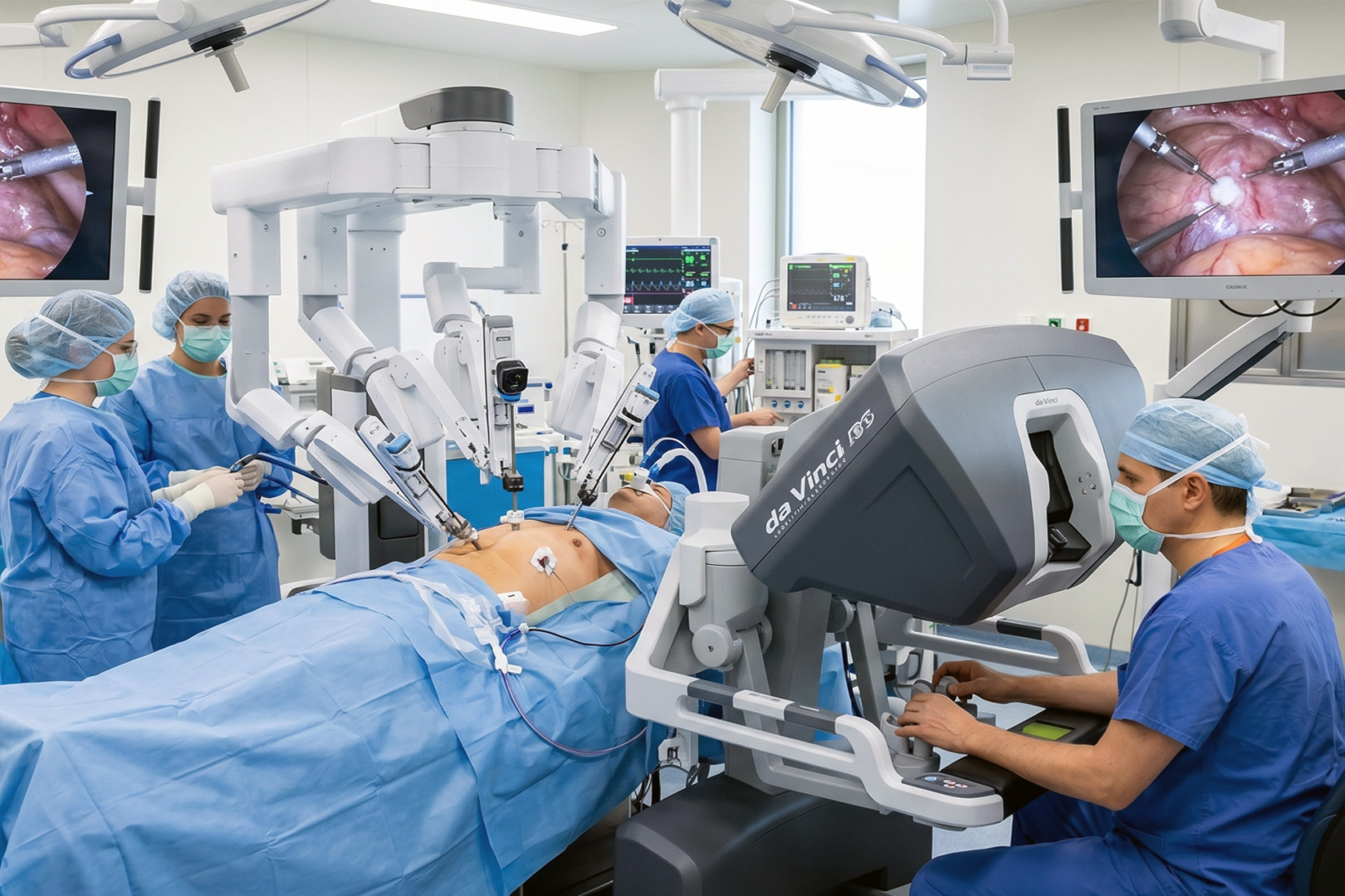
Neumark Lung & Chest Surgery Centre specialises in minimally invasive thoracic surgery with a multidisciplinary approach. Depending on the case, treatment may involve Video-Assisted Thoracoscopic Surgery (VATS), Uniportal VATS, or Robotic Thoracic Surgery (RATS). These techniques are used to remove benign and malignant lung nodules through smaller incisions than traditional open surgery, with the aim of reducing pain and supporting a quicker recovery when appropriate.
Risks and Recovery
Recovery after surgery for lung nodules is often smooth, but every operation still carries risks that should be discussed clearly beforehand.
Possible risks include bleeding, infection, prolonged air leaks, pneumonia, pain and blood clots. The overall risk depends on the type of resection, the patient’s lung function and other medical conditions. After surgery, the removed tissue is examined under the microscope to confirm whether the nodule is benign, pre-cancerous, or malignant, and that result determines whether any further treatment is needed. A biopsy is performed when needed, and cancerous lung nodules are treated promptly.
With minimally invasive approaches, many patients are able to mobilise early and recover faster than with open surgery. Careful pre-operative assessments, including high-resolution CT and selected PET-CT imaging, aid planning, while smaller-incision techniques can facilitate earlier chest drain removal and a smoother post-operative course in suitable patients.

How Neumark Can Help
Neumark Lung & Chest Surgery Centre can help by turning an alarming scan report into a clear, personalised plan. Patients who hear the term ‘spiculated lung nodule’ often fear the worst. What they usually need first is not panic, but a proper interpretation of the scan, a review of any prior imaging, an assessment of risk factors, and a sensible recommendation on whether to watch, biopsy, or remove the nodule.
Neumark provides that stepwise approach through specialist evaluation and minimally invasive thoracic care led by Dr Harish Mithiran, senior consultant thoracic surgeon at Gleneagles and Mount Alvernia hospitals.
If you have been told you have a spiculated nodule, contact Neumark for a consultation.