
Large cell lung carcinoma is an uncommon type of non-small cell lung cancer (NSCLC). Under a microscope, its cells look large and poorly differentiated, but they lack the specific features of adenocarcinoma, squamous cell carcinoma, or small cell lung cancer (SCLC).
A NSCLC tumour can begin in any part of the lung, although it often appears near the outer edges. These tumours may grow and spread quickly. Some people have no symptoms until the cancer is more advanced.
What Is Large Cell Lung Carcinoma?
Non-small cell lung cancer (NSCLC) accounts for about 80% to 85% of lung cancers. Its main types and subtypes include adenocarcinoma, the most common type, squamous cell carcinoma and large cell carcinoma.
Large cell carcinoma is a diagnosis of exclusion within NSCLC. This means the tumour lacks the microscopic features and immunohistochemical markers needed to classify it as a more specific type of lung cancer, so it is a process of elimination. Immunohistochemistry uses laboratory stains to identify the proteins in cancer cells.
In the past, older reports and the World Health Organisation estimated that large cell carcinoma comprised 3% to 9% of lung cancers. Today, it is diagnosed less often under modern classifications because improved testings can identify many previously unclassified tumours as adenocarcinoma, squamous cell carcinoma, or another specific cancer.
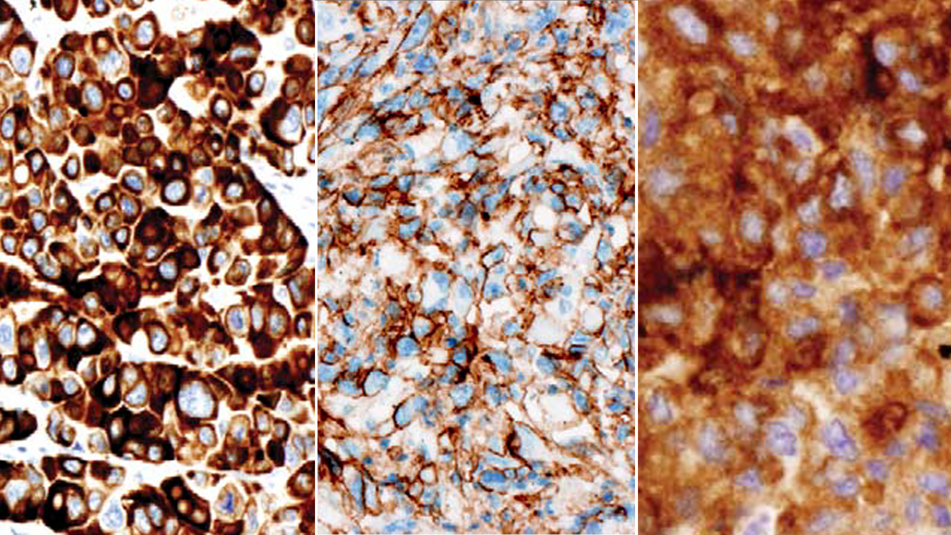
Large Cell Carcinoma Histology
Histology describes how cells and tissue look under a microscope. In large cell carcinoma, the cancer cells are large epithelial cells with abundant cytoplasm and prominent nucleoli.
The pathologist first reviews the cell shape, tissue pattern, and architectural features of the tumour cells. Immunohistochemical stains will then look for evidence of a more specific lineage. For example, TTF-1 and Napsin A may support lung adenocarcinoma, while p40 may support squamous cell carcinoma, which arises from flat cells that line the airways. TTF-1 and Napsin A support lung adenocarcinoma with glandular differentiation, while p40 supports squamous differentiation.
If the tumour shows no glandular or squamous differentiation and no neuroendocrine differentiation after the appropriate testing, it may be classified as a large cell carcinoma. A small biopsy can confirm cancer, but it may not contain enough tissue to exclude every other subtype. In that situation, the initial diagnosis may be non-small cell carcinoma, not otherwise specified.
A definitive diagnosis of large cell carcinoma often requires examination of a larger surgical specimen. Pathologists may describe morphologic variants, such as basaloid carcinoma, lymphoepithelioma-like carcinoma, clear cell carcinoma or a rhabdoid phenotype, though these variants are now often reclassified.
Is Large Cell Neuroendocrine Carcinoma the Same Disease?
No. Older sources sometimes describe large cell neuroendocrine carcinoma (LCNEC) as a subtype of large cell carcinoma. Current classification places LCNEC with high-grade neuroendocrine carcinomas, together with small cell carcinoma and small cell lung cancer, rather than with conventional large cell carcinoma.
LCNEC has neuroendocrine cell patterns, a high rate of cell division and neuroendocrine marker expression. Similar to small cell lung cancer, it is a distinct diagnosis that may require different types of treatment.
Symptoms of Large Cell Lung Carcinoma
Large cell lung cancer does not have a unique symptom pattern. Symptoms may appear late, particularly when a tumour begins in the outer lung and does not obstruct a large airway.
Possible symptoms include:
- A persistent cough or a change in a long-standing cough
- Shortness of breath
- Chest pain, chest wall discomfort or pressure
- Fatigue or weakness
- Unexplained weight loss or a reduced appetite
- Coughing up blood or blood-stained mucus
- Wheezing, hoarseness or repeated chest infections
These symptoms have many possible causes and do not confirm lung cancer. However, persistent, worsening or unexplained symptoms will need an urgent medical assessment, especially for more than a small amount of coughed-up blood, severe or rapidly worsening breathlessness, fainting or sudden chest pains.

Causes and Risk Factors
Cigarette smoking is the leading risk factor for large cell lung cancer and other forms of lung cancer. The risks generally increase with the amount smoked and the number of years a person has smoked. Large cell carcinoma can also occur in people who have never smoked.
Other lung cancer risk factors include secondhand smoke, radon, asbestos, certain workplace carcinogens, outdoor air pollution, previous radiation therapy to the chest, such as after breast cancer, and a personal or family history of lung cancer. Having a risk factor does not mean that cancer will develop, but it increases the chances and requires more vigilance for any unusual symptoms.
How Large Cell Lung Carcinoma Is Diagnosed
The diagnosis will aim to confirm the cancer type, establish its stage and determine which treatment options may be suitable. Blood tests may assess your general health before treatment.
Imaging Tests
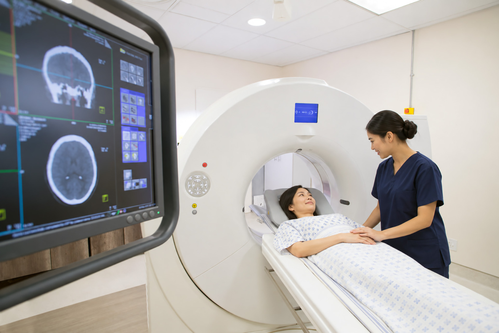
A chest X-ray may first show an abnormal area. A contrast-enhanced CT scan provides more detail about the tumour’s size, location and its relationship to the nearby structures. PET-CT scans may help assess the lymph nodes and the possible spread of the cancer elsewhere. A brain MRI may be recommended depending on the stage, symptoms and the planned treatment.
Imaging can show where a tumour is and how far it may have spread, but it cannot confirm large cell carcinoma. Tissue testing is required for this.
Biopsy and Pathology Review
A lung biopsy removes cells or tissue for laboratory examination. The approach may involve a bronchoscopy, a CT-guided needle biopsy, endobronchial ultrasound or surgery, depending on the tumour’s location and the information needed.
The pathologist will review the tissue and will use immunohistochemical stains to distinguish large cell carcinoma from other types of lung cancer. A second pathology review is useful when the diagnosis remains uncertain, or the tissue sample is limited.
Molecular and Biomarker Testing
Molecular testing looks for changes in genes or proteins in the tumour cells that may influence the treatment. Testing may include a broad genomic panel and PD-L1 assessment, particularly for advanced disease or when the tumour’s lineage is uncertain. An actionable biomarker may make targeted therapy suitable, and PD-L1 results may help guide immunotherapy.
After diagnosis, the healthcare team uses the lung cancer stage to describe the tumour, regional lymph nodes and distant spread. This information, especially on the subtype and stage, affects treatment planning.
Large Cell Lung Carcinoma Treatment
Large cell lung carcinoma treatment generally follows the same principles and options as non-small cell lung cancer (NSCLC). The NSCLC plan depends on stage, tumour location, lymph node findings, molecular and PD-L1 results, lung function, other health conditions, and the person’s preferences.
Surgery for Early-Stage Cancer
Surgery for non-small cell lung cancer is commonly considered for operable Stage I or Stage II diseases. Selected people with resectable Stage III disease may also have surgery as part of a combined treatment plan.
A lobectomy removes one lung lobe and is appropriate for many operable tumours. A segmentectomy or wedge resection may suit selected small tumours or people with limited lung reserve. A pneumonectomy removes an entire lung and is needed less often.
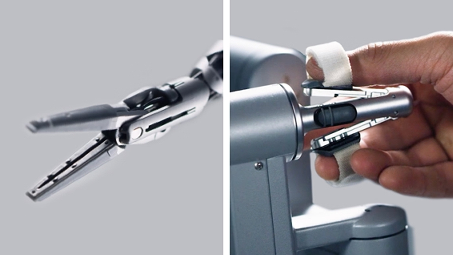
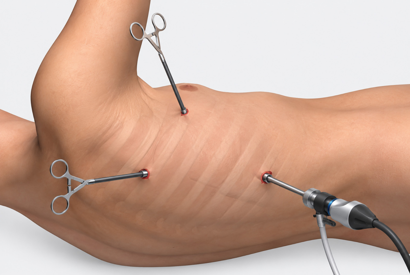
Suitable operations may be performed using Video-Assisted Thoracoscopic Surgery (VATS) or Robotic-Assisted Thoracic Surgery (RATS). These approaches use smaller incisions than open thoracotomy. The appropriate approach ultimately depends on the tumour’s size, position and involvement of nearby structures.
Treatment Before or After Surgery
In NSCLC, chemotherapy, immunotherapy or chemoradiotherapy may be used before surgery in selected cases, to reduce the size of the cancer. Adjuvant chemotherapy may also follow surgery to reduce the risk of recurrence, depending on the pathological stage and other findings. Chemotherapy side effects can include fatigue and hair loss. Postoperative immunotherapy, targeted therapy, or radiation therapy may be appropriate for certain patients, but these therapies are not required for all patients after every operation.
When Surgery Is Not Suitable
Radiation therapy may treat a localised non-small cell lung cancer tumour when surgery is not suitable. Unresectable locally advanced disease often requires a combination of chemotherapy and radiotherapy, sometimes followed by immunotherapy.
For metastatic disease, systemic treatment is usually the main approach. Treatment options may include chemotherapy, immunotherapy, targeted therapy or a combination of these. Targeted therapy uses medicines directed at specific tumour biomarkers, and these therapies vary based on the biomarker found. Immunotherapy helps the immune system recognise or attack cancer cells. The complete lung cancer diagnosis and treatment guide explains these pathways in more detail.
Prognosis and Follow-Up
The outlook depends most strongly on the stage at diagnosis. Tumour location, lymph node involvement, distant spread, biomarkers, general health, ability to completely remove the cancer, and response to treatment also matter.
No single large-cell lung cancer survival rate applies to all situations. Older statistics may include other tumour types that would now receive a different diagnosis than large-cell lung cancer. Advanced disease is less often curable, but treatment may control the cancer and ease symptoms in most patients.
Follow-ups after treatment may include clinical reviews and scheduled CT scans. Report new or worsening symptoms rather than waiting until the next planned appointment.
Thoracic Surgical Assessments at Neumark
Neumark Lung & Chest Surgery Centre specialises in minimally invasive thoracic surgery with a multidisciplinary approach led by Dr Harish Mithiran, senior consultant thoracic surgeon at Gleneagles and Mount Alvernia hospitals.
A thoracic surgical assessment may include a review of CT and PET-CT images, pathology, lymph node findings, molecular results, lung function, and assessments of whether the tumour can be removed completely while preserving sufficient working lung tissue. Care will be coordinated with respiratory physicians, medical and radiation oncologists, radiologists and pathologists.
Patients can contact Neumark to discuss whether a thoracic surgical assessment is appropriate for their lung cancer.
FAQs about Large Cell Lung Carcinoma
Large Cell Carcinoma vs Adenocarcinoma
These are different NSCLC subtypes. Lung adenocarcinoma shows glandular differentiation or markers that support that diagnosis. Large cell carcinoma lacks the defining features of adenocarcinoma, squamous cell carcinoma and other more specific lung cancer types.
Large Cell Carcinoma vs Small Cell Carcinoma
Large cell carcinoma is a type of non-small cell lung cancer. Small cell lung cancer is a separate high-grade neuroendocrine cancer with different pathological features, usual behaviour and treatment.
Can a Small Biopsy Confirm Large Cell Carcinoma?
A small biopsy may confirm non-small cell lung cancer, but it may not provide enough tissue to rule out every more specific subtype. Definitive classification as conventional large cell carcinoma often requires a larger resection specimen.
Is Large Cell Lung Carcinoma Curable?
Some localised cancers can be treated with curative intent, often with surgery and, when indicated, additional therapy. The likelihood of cure decreases after extensive lymph node involvement or distant spread.