
Squamous cell lung carcinoma is a type of non-small cell lung cancer (NSCLC). It usually begins in the larger airways near the centre of the lung and has a strong association with tobacco smoking. Early instances may cause no symptoms, while a central tumour may cause a persistent cough, coughing up blood, or repeated chest infections.
A CT scan can show the tumour and possible airway blockage, but an accurate diagnosis requires a biopsy. The stage, tumour location, nodal findings, lung function, molecular results, and overall health then help guide treatment, whether by surgery or otherwise.
| Feature | Squamous Cell Lung Carcinoma |
| Main classification | Non-small cell lung cancer (NSCLC) |
| Cells involved | Squamous cells that develop from the lining of the airways |
| Usual location | Often central, near a main or lobar bronchus, although peripheral tumours also occur |
| Main preventable risk factor | Tobacco smoking |
| Possible scan features | A central mass, airway obstruction, collapsed lung tissue, consolidation, or a cavity within the tumour |
| How diagnosis is confirmed | Biopsy with microscopic examination and, when needed, immunohistochemistry |
| What guides treatment | Stage, resectability, lung function, pathology, biomarkers and the patient’s overall health |
What Is Squamous Cell Lung Carcinoma?
Squamous cells are flat cells that protect some body surfaces and mucous membranes. They are not normally the main cells lining the bronchi, which are large tubes that carry air from the trachea to the lungs. Repeated irritation from tobacco smoke can cause the airway lining to change into squamous cells, a process called squamous metaplasia. Further DNA changes may allow these cells to grow without normal control. This can lead to dysplasia, carcinoma in situ, and eventually invasive squamous cell carcinoma.
Squamous cell lung carcinoma belongs to the non-small cell lung cancer group. It is different from small cell lung cancer, which is a high-grade neuroendocrine cancer that usually grows and spreads more quickly.
The name describes the cell type, not the stage of the cancer. The disease can range from a non-invasive lesions confined within the airway lining to cancer that has spread to the lymph nodes or distant organs.
Squamous Cell Carcinoma of the Lung Is Not Skin Cancer
Squamous cell carcinoma can begin in several organs, including the skin, lung, mouth, throat, oesophagus and cervix. These cancers share some microscopic features but have different causes, tests, staging systems and treatments.
Cutaneous squamous cell carcinoma, also called squamous cell skin cancer, begins in skin cells and often affects sun-exposed skin after ultraviolet radiation. It is a form of nonmelanoma skin cancer. Basal cell carcinoma is another common skin cancer. Though they may sound like similar names, these forms of skin cancers are different from primary squamous cell carcinomas of the lung.
Primary squamous cell lung carcinoma originates in the lungs, is not caused by ultraviolet light, and does not present as a changing mole. Furthermore, localised skin interventions such as routine skin examinations, Mohs micrographic surgery, topical medicines, and photodynamic therapy play no role in diagnosing or treating primary lung tumours and are only helpful for skin cancers.
However, primary lung cancer can metastasise to the skin in rare cases, where it can clinically manifest as a firm skin bump or nodule, which occasionally serves as the first visible sign of the disease. In these instances, the carcinoma is still treated in the lungs because the lungs are the source of the condition.
If a person has had squamous cancer elsewhere, the team may need to determine whether a lung lesion is a new primary cancer or a metastasis.
Squamous Cell Carcinoma Lung Histology and Pathology
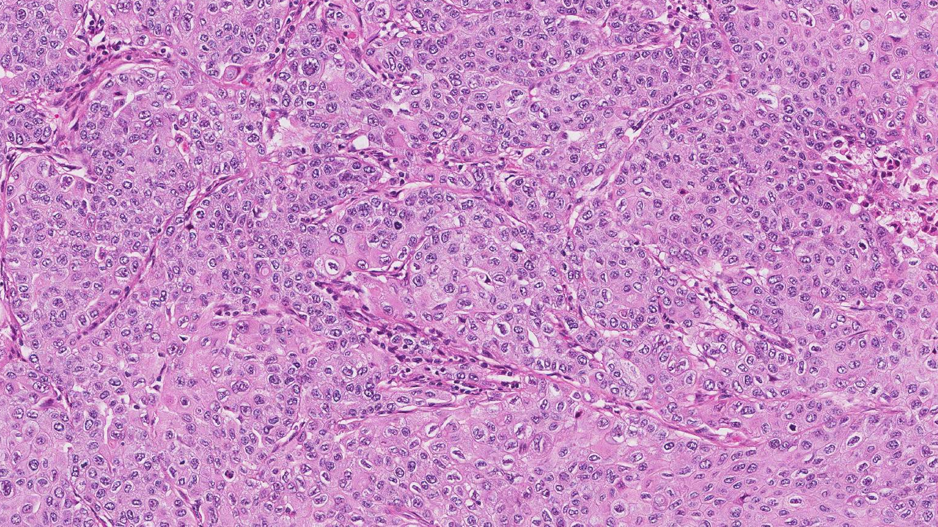
Squamous cell carcinoma lung histology, which describes how cells and tissues look under a microscope, may show keratinisation, in which cancer cells produce keratin, or intercellular bridges between neighbouring cells.
Current cancer pathology classification recognises:
- Squamous cell carcinoma in situ: Abnormal cells remain within the airway lining and do not invade deeper tissue.
- Keratinising squamous cell carcinoma: The invasive tumour forms visible keratin.
- Non-keratinising squamous cell carcinoma: The cancer lacks obvious keratinisation and usually needs immunohistochemistry to confirm squamous differentiation.
- Basaloid squamous cell carcinoma: The tumour has a distinctive basaloid pattern under the microscope.
Lung Squamous Cell Carcinoma in Situ
Lung squamous cell carcinoma in situ is confined to the airway lining. It may be difficult to see on a standard CT scan because it can grow along the bronchial surface without forming a substantial mass. Bronchoscopy may identify and sample the abnormal area. Treatment management will depend on its site and extent, airway anatomy, other lung diseases and whether an invasive cancer is present.
Immunohistochemistry
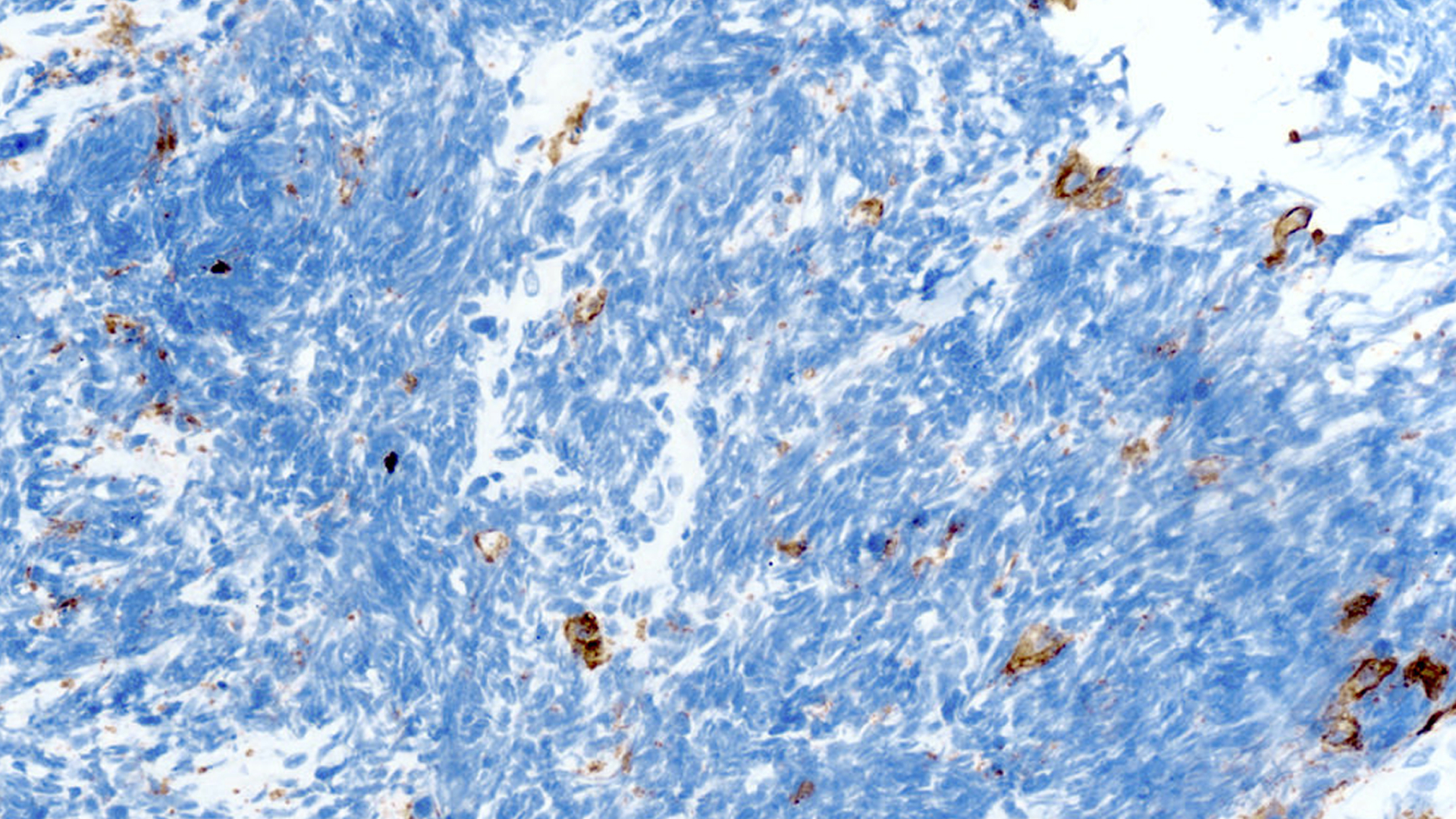
On a small biopsy, cancer features may not be visible. A pathologist may use immunohistochemistry, which applies laboratory stains to detect proteins in the cancer cells.
Squamous tumours commonly express p40 and cytokeratin 5/6; p63 may also be positive but is less specific. Markers associated with lung adenocarcinoma, such as TTF-1 and Napsin A, are usually absent. The pathologist considers the cell appearance, staining pattern, imaging and medical history together.
Adenocarcinoma vs Squamous Cell Carcinoma Lung Cancer
Lung adenocarcinoma and squamous cell carcinoma are both forms of NSCLC. Imaging tests may suggest one pattern, but only tissue testing can establish the diagnosis.
| Feature | Squamous Cell Lung Carcinoma | Lung Adenocarcinoma |
| Usual location | Often central, near larger bronchi | Often peripheral, in the outer lung |
| Smoking association | Strong | Can occur with or without a smoking history |
| Airway effects | May obstruct a bronchus, cause bleeding or lead to post-obstructive infection | May remain silent while small and peripheral |
| Cavitation | More characteristic | Can occur but is less characteristic |
| Actionable molecular alterations | Identified less often | Identified more often |
| Common pathological markers | p40 and cytokeratin 5/6 | TTF-1 and Napsin A |
These are general patterns. Some squamous tumours arise in the outer lung, and smoking history alone cannot establish the cell type.
Small Cell Lung Cancer vs Squamous Cell Carcinoma
These are separate diseases. Small cell lung cancer consists of neuroendocrine cancer cells, often spreads early and is usually treated mainly with systemic therapy and radiation therapy to contain the spread. Squamous cell lung carcinoma is a NSCLC, and surgery is more commonly considered when it remains localised, as it is simpler to extract with fewer potential complications.
Symptoms of Squamous Cell Lung Carcinoma
Early squamous cell lung carcinoma may cause no symptoms. When the tumour begins near a central airway, symptoms can arise from irritation, bleeding or blockage of the bronchus.
Possible lung cancer symptoms include:
- A cough that does not go away or changes from its usual pattern
- Coughing up blood or blood-streaked mucus
- Shortness of breath, wheezing or noisy breathing
- Chest, shoulder or upper-back pain
- A hoarse voice
- Repeated pneumonia or bronchitis in the same part of the lung
- Fevers that accompany a post-obstructive infection
- Unexplained weight loss, reduced appetite or persistent tiredness
Seek prompt medical assessment for any coughing up of blood. Additionally, seek emergency care if there is more than a small amount of blood, the bleeding does not stop, or it occurs with severe breathlessness, chest pain, faintness or a rapid heartbeat. New weaknesses, confusion, a seizure, or severe breathlessness also needs urgent assessment from a medical professional.
Squamous Cell Carcinoma, Hypercalcaemia and Paraneoplastic Effects
Squamous cell lung carcinoma can release parathyroid hormone-related protein, or PTHrP. This may raise the blood calcium level, called hypercalcaemia. This is a paraneoplastic effect, so the symptoms arise from substances produced by the tumour rather than a direct growth.
Hypercalcaemia may cause thirst, frequent urination, constipation, nausea, weakness, drowsiness, confusion or kidney problems. Markedly high calcium needs prompt medical care, as it can have other causes.
Causes and Risk Factors
Tobacco smoking is the main risk factor for squamous cell carcinoma. The risks generally rise with the number of cigarettes smoked and the years of smoking. Stopping lowers the risk over time and may improve the general health and the ability to tolerate treatment.
Other recognised lung cancer risk factors include:
- Secondhand tobacco smoke
- Radon
- Occupational exposure to asbestos, silica, arsenic, chromium, nickel or diesel exhaust
- Outdoor air pollution
- Previous radiation therapy to the chest
- A personal or family history of lung cancer
- Chronic lung disease in some people
Squamous cell lung cancer can also occur in a never-smoker, but this is less common than lung adenocarcinoma. Risk factors cannot confirm or exclude the diagnosis.
How Squamous Cell Lung Carcinoma Is Diagnosed
A squamous cell carcinoma diagnosis requires a biopsy to confirm the cancer type. Further assessments establish the stage, determine whether the tumour can be removed completely, and help assess the patient’s fitness for treatment.
Imaging
A chest X-ray may show a mass, collapsed lung tissue or infection, but it can miss smaller tumours. A contrast-enhanced chest CT will show the tumour, airways, lymph nodes and nearby structures in greater detail.
The scan may show a central mass that narrows a bronchus, sometimes with a cavity caused by a tumour breakdown. Other findings can include atelectasis, post-obstructive consolidation, enlarged nodes or fluid around the lung. However, these features are not enough for a diagnosis on their own.
PET-CT scans may help assess the lymph nodes and the possible spread beyond the primary tumour. A brain MRI may be recommended depending on the clinical stage, symptoms, and the planned treatment.
Low-dose CT is a lung cancer screening test for selected people at high risk who do not have symptoms. It is not the appropriate test for investigating someone who already has symptoms or an abnormal scan.
Biopsy and Lymph-Node Assessment
A lung cancer biopsy provides cells or tissue for examination. The biopsy route depends on the tumour’s position and the information needed.
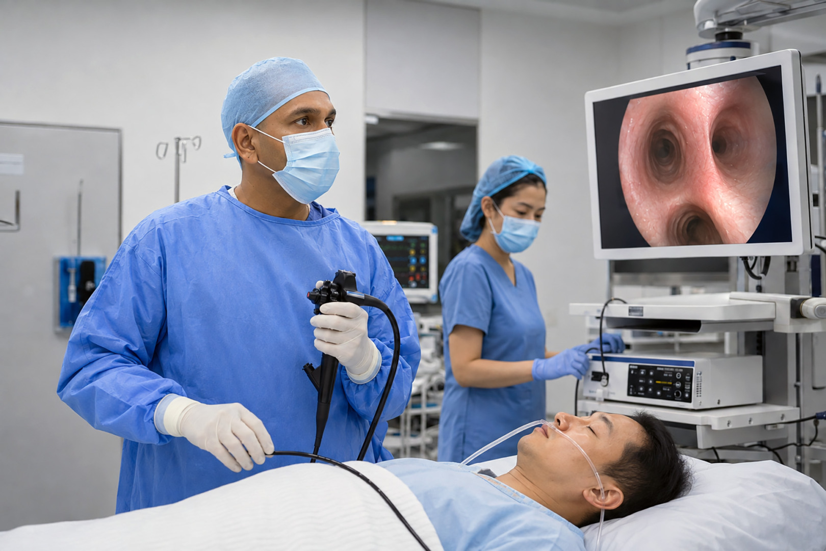
As squamous tumours often arise near a large airway, a bronchoscopy may allow the doctor to see and sample the lesion. An endobronchial ultrasound (EBUS) can sample the central chest lymph nodes. A CT-guided needle biopsy may suit a peripheral tumour. A thoracoscopy or another surgical biopsy is sometimes needed, depending on the situation.
Squamous Cell Carcinoma Mutations and Biomarkers
Squamous cell carcinoma mutations often reflect tobacco-related DNA damage. Changes involving tumour-suppressor pathways, including TP53 and CDKN2A, are common. Alterations with the established targeted treatments occur less often than in lung adenocarcinoma.
For advanced diseases, the team may assess the PD-L1 expression and use molecular profiling when the results may change the treatment. Broader testing can be especially relevant for a never-smoker, a younger patient, a small biopsy with uncertain subtype, or a tumour with mixed features.
Staging
Squamous NSCLC uses the TNM staging system, which considers the primary Tumour, regional lymph Nodes, and distant Metastases. The lung cancer stages range from Stage 0 to Stage IV.
- Stage 0 describes squamous carcinoma in situ, where the carcinoma is limited to the original site.
- Stage I usually describes cancer confined to the lung without nodal involvement.
- Stages II and III reflect increasing local invasion or regional spread.
- Stage IV includes a separate tumour nodule in the opposite lung, pleural or pericardial tumour deposits, a malignant pleural or pericardial effusion, or distant spread.
Squamous Cell Carcinoma Lung Treatment
Squamous cell carcinoma lung treatments depend on the stage, tumour location, nodal findings, molecular and PD-L1 results, lung function, other medical conditions and the patient’s preferences.
Surgery for Early-Stage Disease
Lung cancer surgery is commonly considered for operable Stage I or II squamous NSCLC. Selected patients with resectable Stage III disease may also have surgery as one part of combined treatment after multidisciplinary review.
The operation may include:
- Lobectomy: Removes one lobe and remains appropriate for many operable tumours.
- Segmentectomy: Removes an anatomical lung segment and may suit selected small peripheral tumours.
- Sleeve resection: Removes a tumour-bearing section of bronchus and reconnects the airway. For a suitable central tumour, this can avoid removing the entire lung.
- Pneumonectomy: Removes one lung that may be necessary for some central tumours.
- Wedge resection: Removes the tumour with a small rim of lung and is reserved for selected situations.
Lymph-node sampling or dissection forms an important part of surgery. It confirms the pathological stage and helps guide post-operative treatment.
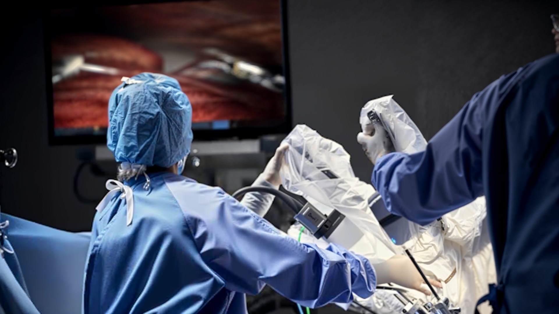
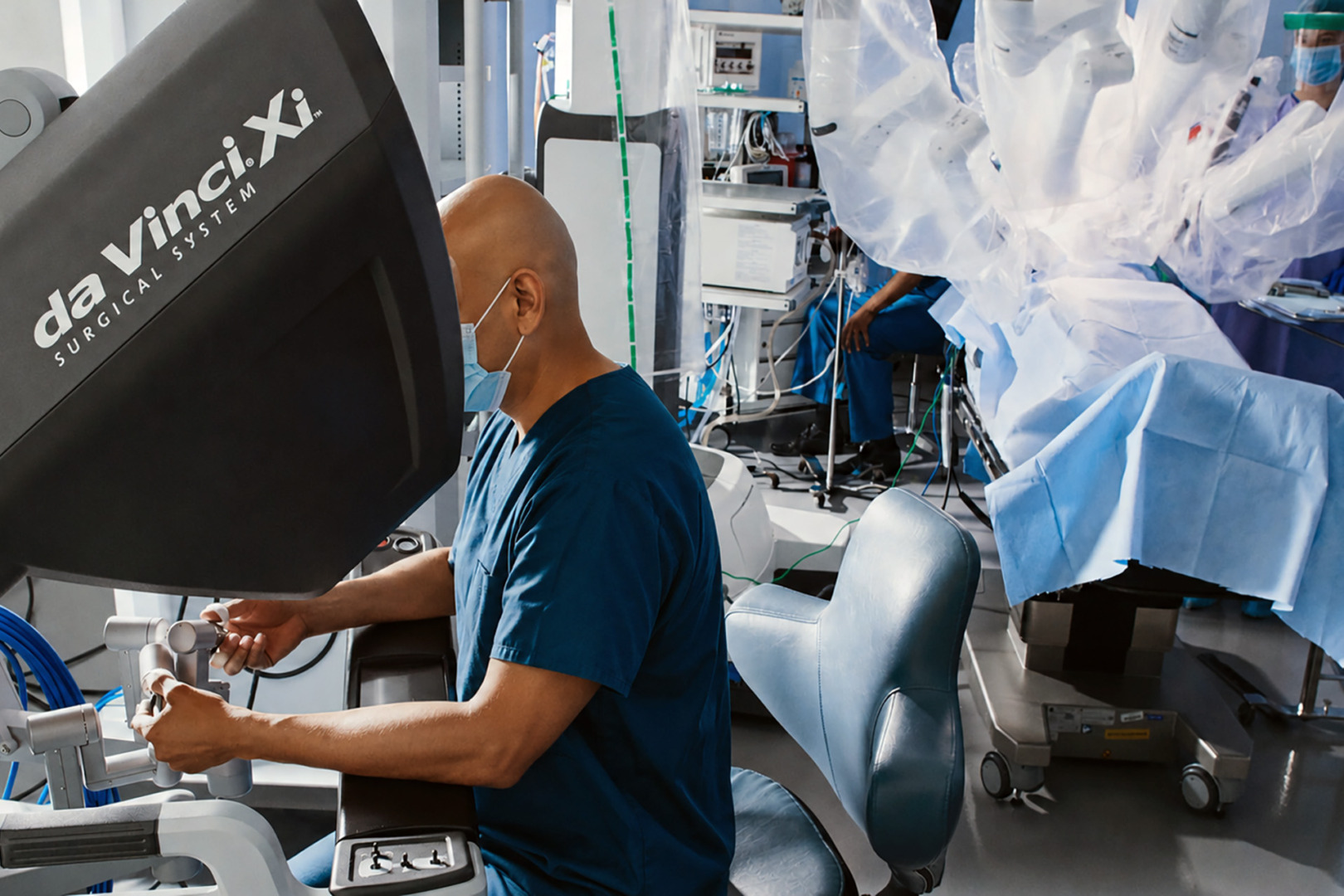
Video-Assisted Thoracoscopic Surgery (VATS), including Uniportal VATS, and Robotic-Assisted Thoracic Surgery may treat squamous cell carcinoma in suitable cases. These approaches use smaller incisions than a thoracotomy, but they are not appropriate for every squamous cancer. A tumour involving a main airway, major blood vessel or nearby structure may require an invasive operation.
Treatment Before or After Surgery
Lung cancer treatment before surgery may include chemotherapy with immunotherapy, chemotherapy alone, or chemoradiotherapy in selected cases. After surgery, doctors may consider chemotherapy or immunotherapy based on the pathological stage, lymph nodes, margins, and biomarker results. Postoperative radiation therapy is reserved for particular findings. These treatments are not required after every operation.
When Surgery Is Not Suitable
Stereotactic body radiation therapy may be considered for a localised tumour when a patient cannot have surgery, due to their suitability for an operation. Central tumours require careful radiation planning because the larger airways, heart, oesophagus and major blood vessels lie nearby.
For many with unresectable, locally advanced disease, combined chemotherapy and radiation therapy forms the main treatment. Immunotherapy may follow in suitable cases.
Squamous Cell Carcinoma Lung Cancer Stage 4
Stage 4 is when squamous cell carcinoma lung cancer has spread to the opposite lung, pleura, pericardium, other organs, or another distant site. Systemic treatment is usually the main approach rather than exclusively surgery at this stage.
Options may include immunotherapy, chemotherapy or both. Immunotherapy can help the immune system recognise and destroy cancer cells. Molecular results, PD-L1 expression, symptoms, previous treatment and overall health help guide the plan. Radiation therapy or another local treatment may help with pain, bleeding, airway obstruction or selected areas of spread.
Surgery does not usually treat widespread Stage IV disease. It may have a limited role in highly selected situations or to manage a complication. Supportive and palliative care can continue alongside cancer-directed treatment.
Prognosis and Follow-Ups
The outlook depends most strongly on the cancer stage. Tumour size and location, nodal involvement, distant spread, ability to remove the cancer completely, the patient’s overall health, and the response to treatment.
Statistics for cutaneous squamous cell carcinoma do not apply to lung cancer. No single squamous cell lung carcinoma survival rate can predict what will happen to one person. Population figures combine all NSCLC subtypes, use older staging systems or reflect treatments used several years earlier.
Some localised cancers can be treated with curative intent, particularly when diagnosed and treated early. At an advanced stage, a complete cure is less likely, but treatment may control the cancer and ease symptoms. The healthcare team can explain the prognosis based on the individual’s stage, test results, and response.
Follow-up after treatment may include clinical review and scheduled chest CT scans. The timing will depend on the stage and treatment received. Report new coughing up of blood, worsening breathlessness, unexplained weight loss, neurological symptoms, or persistent bone pain rather than waiting until the next planned visit.
Thoracic Surgical Assessment at Neumark
Neumark Lung & Chest Surgery Centre specialises in minimally invasive thoracic surgery with a multidisciplinary approach led by Dr Harish Mithiran, senior consultant thoracic surgeon at Gleneagles and Mount Alvernia hospitals.
A thoracic surgical assessment may include CT and PET-CT images, pathology, nodal findings, lung function and whether the tumour can be removed with clear margins while preserving sufficient lung. Care may be coordinated with respiratory physicians, oncologists, radiologists and pathologists.
Patients can contact Neumark to discuss whether a thoracic surgical assessment is appropriate.
FAQs about Squamous Cell Lung Carcinoma
Is Squamous Cell Lung Carcinoma Curable?
Some localised cancers can be treated with curative intent, often with surgery or focused radiation therapy. The chance of cure becomes lower after an extensive nodal involvement or distant spread of the cancer.
Does Squamous Cell Lung Carcinoma Always Start Centrally?
No. It often begins near a larger bronchus, but peripheral tumours also occur. Imaging cannot confirm the cell type.
Can a Never-Smoker Develop Squamous Cell Lung Carcinoma?
Yes, but it is less common. Tobacco exposure has a stronger association with this subtype than with lung adenocarcinoma. Some people have no clear cause.
Is Squamous Cell Lung Carcinoma the Same as Small Cell Lung Cancer?
No. Squamous cell carcinoma is a type of NSCLC. Small cell lung cancer is a separate neuroendocrine cancer that usually behaves and is treated differently.
What Does a Cavitary Squamous Lung Tumour Mean?
A cavity is an air-filled or fluid-filled space within a tumour, often caused by tissue breakdown. Infection and other conditions can look similar, so a biopsy is needed.