
Lung adenocarcinoma is the most common type of lung cancer. It is a form of non-small cell lung cancer (NSCLC) that shows gland-forming features, or produces mucin, a substance found in mucus. It often begins in the outer parts of the lung, but it can spread.
Lung adenocarcinoma rates are particularly high in Southeast Asia. It can affect current smokers, former smokers and never-smokers and is the predominant lung cancer subtype among never-smokers. It is more common for women than men and can occur in younger adults, although lung cancer overall is diagnosed mainly in older people.
Early lung adenocarcinoma may not cause symptoms. A CT scan may first show it as a solid, part-solid or ground-glass lung nodule. Imaging can raise suspicion, but a tissue sample is usually needed to confirm a lung adenocarcinoma diagnosis. Stage, microscopic pattern, molecular features, and overall health then help guide the treatment plan.
What Is Lung Adenocarcinoma?
Lung adenocarcinoma begins when epithelial cells in the lung develop changes that allow them to grow without normal control. Under a microscope, the cancer cells form gland-like structures, produce mucin, or show both features.
It belongs to the broader non-small cell lung cancer group. NSCLC generally grows and spreads less quickly than small cell lung cancer (SCLC), although the behaviour of an individual non-small cell lung adenocarcinoma can vary considerably.
| Feature | Lung Adenocarcinoma |
| Main classification | Non-small cell lung cancer (NSCLC) |
| Usual site | Often in peripheral lung tissue, away from the larger central airways |
| Possible scan appearance | Solid, part-solid or ground-glass nodule, mass or persistent area of consolidation |
| How it is confirmed | Pathological examination of a biopsy or surgical specimen |
| What guides treatment | Stage, resectability, histological pattern, molecular results, PD-L1 expression and overall health |
The term adenocarcinoma describes the tumour’s cell type. It does not describe its stage. Lung adenocarcinoma can range from a small non-invasive lesion to cancer that has spread to the lymph nodes or distant organs.
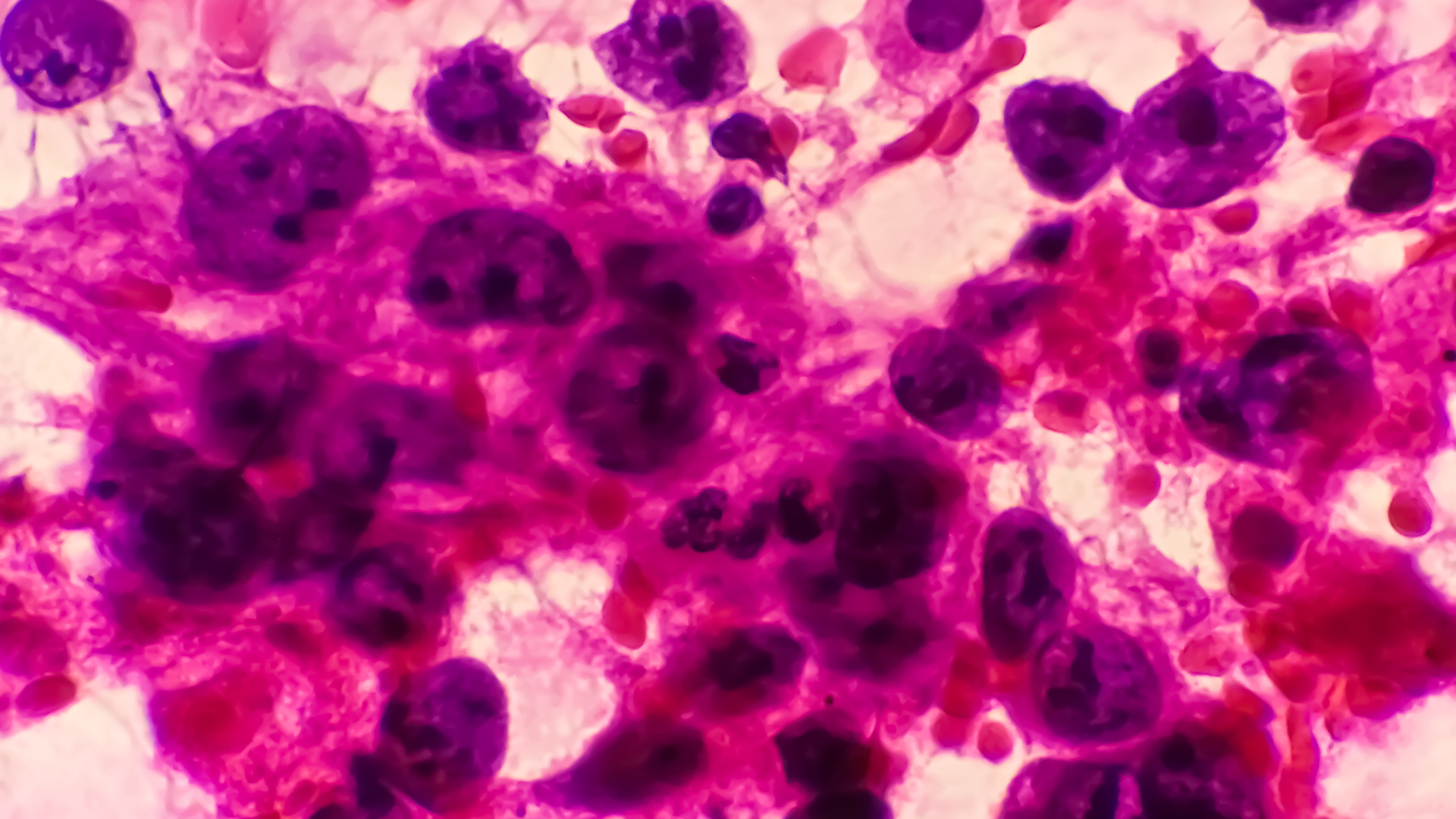
Lung Adenocarcinoma Histology and Subtypes
Lung adenocarcinoma histology describes how the tumour cells grow and look under a microscope. This matters because tumours within the adenocarcinoma family do not all behave the same way.
The main pathological categories include:
- Adenocarcinoma in situ (AIS). This small, non-invasive tumour grows along existing air sacs. Adenocarcinoma in situ measures no more than 3 centimetres with pure lepidic growth and no stromal, vascular or pleural invasion.
- Minimally invasive adenocarcinoma (MIA). Minimally invasive lung adenocarcinoma measures no more than 3 centimetres, is predominantly lepidic and has an invasive component measuring no more than 5 millimetres. Definitive classification generally requires examination of the entire tumour.
- Invasive adenocarcinoma. This cancer has entered the supporting lung tissue. The predominant pattern of invasive lung adenocarcinoma may be lepidic, acinar, papillary, micropapillary or solid. Acinar lung adenocarcinoma forms distinct gland-like structures within the acinus.
- Invasive mucinous adenocarcinoma. These tumours contain mucin-producing cells and may appear as a mass, several nodules, or an area of consolidation resembling pneumonia.
Many invasive tumours contain more than one pattern. Micropapillary and solid-predominant patterns tend to have a higher risk of recurrence than lepidic-predominant tumours. Pleural, lymphatic, or blood vessel invasion and lymph node involvement also affect risk assessment.
Adenocarcinoma Versus Squamous Cell Carcinoma of the Lung
Adenocarcinoma and squamous cell lung carcinoma are both forms of NSCLC, but they differ in their usual location, cell features and association with smoking.
| Feature | Lung Adenocarcinoma | Squamous Cell Lung Carcinoma |
| Usual location | Often peripheral | Often near a large central airway |
| Smoking association | Can occur with or without a smoking history | More strongly associated with smoking |
| Prevalence in never-smokers | Most common lung cancer type in never-smokers | Less common in never-smokers |
| Molecular testing | Often identifies alterations that can guide targeted therapy | Actionable alterations are found less often |
| Airway symptoms | May remain silent while small and peripheral | A central tumour may cause cough, airway blockage or coughing up blood |
These are general patterns, not rules. Cases of lung adenocarcinoma can vary widely and overlap with cases of squamous cell carcinoma.

Lung Adenocarcinoma Symptoms
Early lung adenocarcinoma often causes no symptoms. It may be found as a lung nodule during screening or a scan performed for another reason.
When symptoms occur, they may include:
- A chronic cough that does not go away or changes from its usual pattern
- Shortness of breath or wheezing
- Chest, shoulder or upper-back pain
- Coughing up blood
- A hoarse voice
- Repeated or slow-to-resolve chest infections
- Unexplained weight loss or a reduced appetite
- Persistent tiredness
Breathlessness may result from airway narrowings, a collapsed section of the lung, or a malignant pleural effusion. Spreading to the brain may cause headaches, weakness, confusion or seizures, while bone involvement may cause persistent pain.
These lung cancer symptoms and causes can result from many conditions other than cancer. Seek emergency care if you experience severe or sudden shortness of breath, or cough up blood, especially if it is accompanied by chest pain, faintness, or a rapid heartbeat.
Causes and Risk Factors
Lung adenocarcinoma develops when cells acquire DNA changes that disrupt normal growth and repair. These changes usually develop within the tumour during a person’s lifetime and are not necessarily inherited via genetics.
Smoking remains the main preventable risk factor for lung cancer, including adenocarcinoma. The risks rise with the amount and duration of tobacco exposure. Stopping smoking lowers the risk over time, although former smokers remain at increased risks for years. However, lung adenocarcinoma is also known to affect never-smokers.
Other factors associated with lung cancer include:
- Secondhand tobacco smoke
- Radon exposure
- Workplace exposure to asbestos, silica, arsenic, chromium, nickel or diesel exhaust
- Outdoor air pollution
- Previous radiation therapy to the chest
- A family history of lung cancer
- Certain chronic lung diseases, including pulmonary fibrosis
Lung Adenocarcinoma in Never-Smokers
Lung adenocarcinoma is the main lung cancer type found in never-smokers. This is particularly relevant in Singapore and other Southeast Asian populations, where a substantial share of lung cancer occurs without a smoking history.
Never-smokers are more likely to have certain acquired molecular alterations, including changes involving EGFR or ALK. These findings may guide treatment but usually do not mean that the cancer was inherited.
There is no evidence-based recommendation for every never-smoker to have an annual CT scan. In Singapore, routine low-dose CT lung cancer screening is recommended for defined higher-risk groups based mainly on age and smoking history. Importantly, people who have symptoms need diagnostic assessments rather than screening tests.
How Lung Adenocarcinoma Is Diagnosed
The diagnostic process determines whether an abnormality is cancer, identifies its cell type, and establishes how far it has spread.
Imaging
A chest X-ray may show a mass, collapsed lung or fluid around the lung, but it can miss small lesions. A chest CT provides more detail about an abnormality’s size, location, density and borders.
Lung adenocarcinoma can appear as a ground-glass, part-solid or solid nodule, a mass, several pulmonary nodules or persistent consolidation. Infection and non-cancerous conditions can look similar, so CT findings alone will not confirm adenocarcinoma.
PET-CT scans combine anatomical imaging with metabolic activity and may help assess lymph nodes and a possible spread. Some lepidic or mucinous tumours show little uptake, so a low-uptake result does not always rule out cancer. Doctors may recommend brain MRIs based on stage, symptoms, and planned treatment.
Biopsy and Pathology
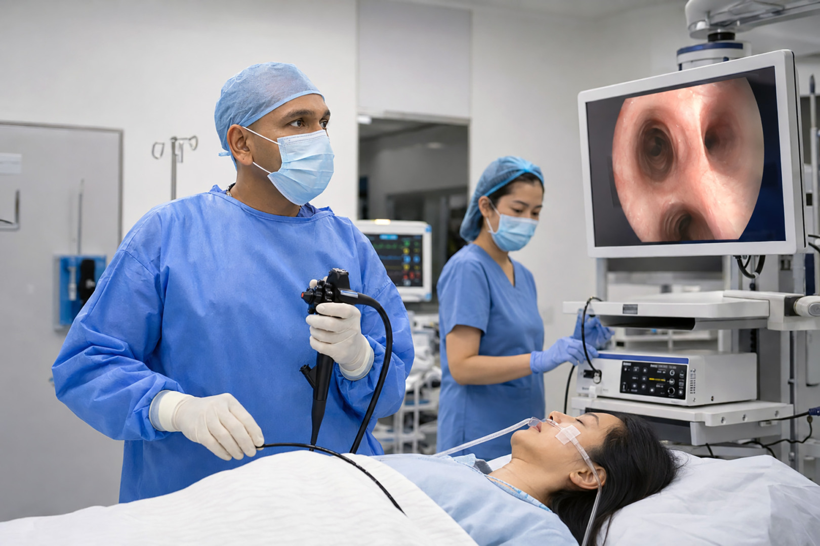
A lung cancer biopsy provides cells or tissue for examination. Depending on the lesion and information needed, the route may include a bronchoscopy, endobronchial ultrasound, CT-guided needle or surgical biopsy.
A pathologist examines the tissue and may use immunohistochemistry. Markers such as TTF-1 and Napsin A are often present, but no single marker proves a definitive diagnosis in every case. The results are interpreted with the tumour’s appearance, imaging findings and clinical history.
Molecular and PD-L1 Testing
Molecular profiling looks for acquired changes within the cancer cells. Depending on the stage and clinical setting, testing may include EGFR, ALK, KRAS, ROS1, BRAF, MET, RET, NTRK and ERBB2, also called HER2. PD-L1 testing measures a protein that can help guide immunotherapy use. PD-L1 is not a gene mutation.
Lung adenocarcinoma mutations vary by ancestry, cigarette smoking habits, stage and testing methods. Figures such as TP53 in 46%, EGFR in 27% or KRAS in 32% describe particular study populations, not universal rates. EGFR alterations are more frequent in Southeast Asian patients and never-smokers, while KRAS alterations are more common in populations with a smoking history.
ALK rearrangements occur in a minority of adenocarcinomas but are enriched in younger patients and never-smokers. EGFR and KRAS driver alterations are usually mutually exclusive, although a rare co-occurrence can happen. TP53 changes are common but do not usually select for a standard targeted therapy on their own.
Broad panel testing may identify an actionable alteration while preserving limited biopsy tissue. A liquid biopsy may detect tumour DNA in the blood, but a negative result does not rule out an alteration, and tissue testing may still be needed.
How Lung Adenocarcinoma Is Staged
Lung adenocarcinoma is staged from Stage 0 to Stage IV using the current TNM system (Tumour-Node-Metastasis). The TNM system assesses the primary lung tumour’s size and growth, the involvement of nearby lymph nodes, and the presence of distant metastases. Lung cancer stages include:
- Stage 0 refers to lung adenocarcinoma in situ, meaning it is at the original site.
- Stage I is generally confined to the lung without lymph-node spread.
- Stages II and III describe increasing local or regional spread.
- Advanced Stage IV includes a separate tumour nodule in the opposite lung, pleural or pericardial nodules, a malignant pleural or pericardial effusion, or metastasis outside the chest.
The number and location of the involved nodes, tumour characteristics, the response to initial treatment, and the patient’s overall fitness all matter. Selected lung cancer cases may be considered for surgery after multidisciplinary reviews, while others are better managed without any operation.
Lung Adenocarcinoma Treatment
Adenocarcinoma lung cancer treatment depends on the stage, resectability, molecular and PD-L1 results, lung function, other health conditions and the person’s preferences.
Early and Locally Advanced Disease
Lung cancer surgery is commonly considered for operable, early-stage lung adenocarcinoma. It may remove a lobe, one or more anatomical segments, a wedge of lung or, less often, an entire lung.
Selected small peripheral tumours may be suitable for segmentectomy. A lobectomy remains appropriate for many operable cancers. The choice depends on tumour size, location, lung function and whether clear margins and adequate lymph-node assessments can be achieved.
Video-Assisted Thoracoscopic Surgery (VATS), including Uniportal VATS, and Robotic-Assisted Thoracic Surgery may be used for suitable tumours. They use smaller incisions than a thoracotomy, but they may not be appropriate for every cancer.
Treatments before surgery may include chemotherapy with immunotherapy, chemotherapy alone or chemoradiotherapy in selected cases. After the surgery, chemotherapy, immunotherapy or eligible targeted therapy may be considered. Postoperative radiotherapy is reserved for specific situations, such as selected nodal disease or involved surgical margins.
If surgery is unsuitable for a localised tumour, stereotactic body radiation therapy may be considered. Locally advanced diseases may require combined chemotherapy and radiation therapy, while selected cases may include surgery.
Advanced or Metastatic Disease
For advanced lung adenocarcinoma, broad molecular testing and PD-L1 assessment should generally be available before selecting the first systemic treatment.
Platinum-based chemotherapy remains important, but it is not the universal first-line of treatment for every advanced adenocarcinoma. An actionable molecular alteration may make targeted therapy appropriate. If no actionable driver is found, treatments may involve immunotherapy, chemotherapy, or both. Radiation or another local treatment may control symptoms or selected areas of spread. Supportive and palliative care can continue alongside cancer-directed treatments.

Lung Adenocarcinoma Prognosis
Lung adenocarcinoma prognosis depends most strongly on the stage at the lung cancer diagnosis. Other factors include the tumour’s predominant growth pattern, lymph-node involvement, molecular features, the patient’s overall health, response to treatment, and whether the surgery has achieved clear margins.
AIS and MIA have an excellent disease-specific outlook after complete resection. Importantly, any lung adenocarcinoma survival rate is a population estimate, not an individual prediction. It is not reliable to apply a single figure to every Stage I or Stage IV cancer case because published percentages may reflect earlier staging editions, different populations, or older treatments.
Follow-up after treatment may include clinical reviews and chest CT scans. The schedule depends on the stage, the treatment received and the risk of recurrence. Report new or worsening symptoms rather than waiting until the next planned doctor’s appointment.
Thoracic Surgical Assessment at Neumark
Neumark Lung & Chest Surgery Centre specialises in minimally invasive thoracic surgery with a multidisciplinary approach led by Dr Harish Mithiran, senior consultant thoracic surgeon at Gleneagles and Mount Alvernia hospitals.
A thoracic surgical assessment may include a review of CT imaging and PET scan, biopsy and molecular results, lymph node findings, lung function and the amount of lung that may need removal. When other treatment is required, care may be coordinated with respiratory physicians, medical oncologists, radiation oncologists, radiologists and pathologists.
Contact Neumark to discuss whether a thoracic surgical assessment is appropriate for you or get guidance on your lung adenocarcinoma.
FAQs about Lung Adenocarcinoma
Is Lung Adenocarcinoma Curable?
Some localised lung adenocarcinomas can be treated with curative intent, often with surgery or focused radiation. A cure becomes less likely after distant spread, but systemic treatment may control the cancer for some patients.
Can a Never-Smoker Develop Lung Adenocarcinoma?
Yes. Adenocarcinoma is the lung cancer type most often diagnosed in never-smokers. Other risk factors may contribute, but in some patients no clear cause can be found.
What Does EGFR-Positive Lung Adenocarcinoma Mean?
It means testing found an acquired EGFR mutation in the cancer cells. The exact alteration matters because some changes can guide targeted therapy. It usually does not mean that there is a genetic link.