Thoracotomy is open chest surgery used when direct access to the lungs or chest organs is needed for treatment or diagnosis.
What Is Thoracotomy?
A thoracotomy is a major open-chest surgery that provides a surgeon with direct access to the lungs, pleura, oesophagus, diaphragm and other structures within the chest. It may be done to remove diseased tissue, repair injury, drain an infection, take a biopsy, or perform major chest surgery, usually under general anaesthesia.
A thoracotomy is a major surgical procedure, so surgeons only recommend it when the expected benefit outweighs the risks or when open access is the safest way to treat the condition. During the surgery, the surgeon makes a skin incision in the chest wall, usually between the ribs, that passes through subcutaneous tissue and intercostal muscles. This creates direct access to the chest cavity.
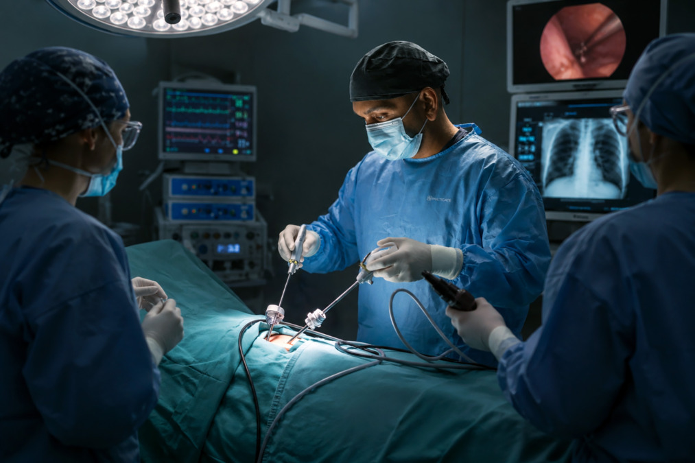
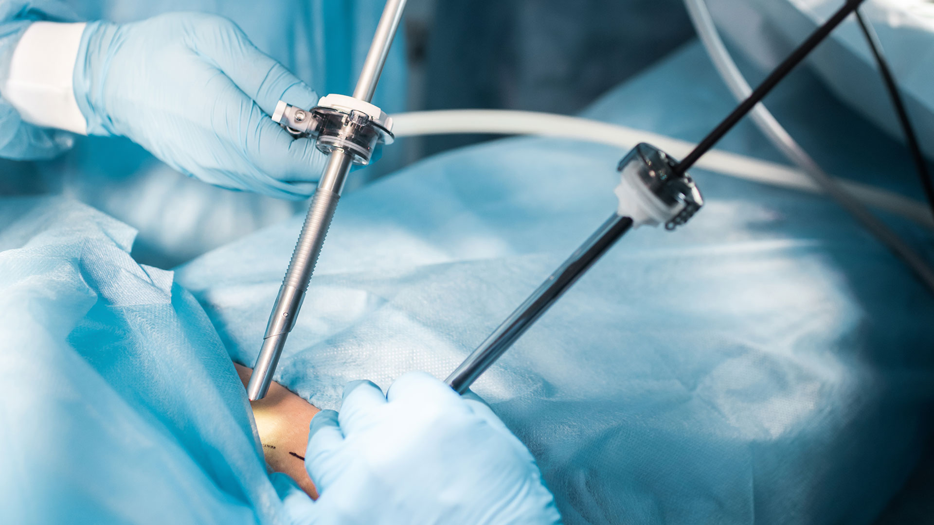
A chest thoracotomy is different from minimally invasive thoracic surgery. In minimally invasive surgery, smaller incisions and a camera are used. In an open thoracotomy, the incision is larger because the surgeon needs wider access.
Schedule a Consultation
What Conditions Does a Thoracotomy Treat?
A thoracotomy may be used to diagnose, treat or control serious conditions affecting the lungs, pleura and other organs in the chest. In lung and pleural disease, it may be considered when minimally invasive surgery is not suitable, or when the condition is too complex for smaller incisions.
A thoracotomy may also be needed for lung removal surgery. This can include:
A wedge resection removes a small piece of lung
A segmentectomy removes a larger anatomical section
A lobectomy removes one lobe of the lung
A pneumonectomy removes an entire lung
Cancer Care
In lung cancer care, a thoracotomy may be used to remove the disease, confirm a diagnosis, stage the disease or relieve symptoms in adults and paediatric patients. It does not always cure cancer. The role of lung cancer surgery depends on the cancer type, stage, patient fitness, lung function and whether other treatment such as chemotherapy, immunotherapy, targeted therapy or radiation therapy is needed.
Emergency Room Thoracotomy
An emergency thoracotomy is different from a planned thoracotomy. It is an operation used after severe chest trauma, such as a major chest injury, heavy bleeding, or a cardiac arrest after a penetrating injury.
It may also be needed if blood or fluid builds up around the heart and stops it from filling properly. This is called a cardiac tamponade and may be seen on an urgent bedside ultrasound scan.
In trauma care, an emergency thoracotomy may be considered if a patient has no pulse shortly after an injury, if blood pressure remains dangerously low despite resuscitation, or if there is a rapid bleeding from a chest tube. In these situations, the aim is to control bleeding, relieve pressure around the heart, and give the patient the best chance of survival.
Schedule a Consultation
When to See a Thoracic Surgeon
You should see a thoracic surgeon when your chest condition may need a biopsy, surgical treatment, or direct access to the lungs or pleural space.
Specialist review is important if imaging shows a lung or pleural tumour, a trapped lung, a persistent pleural infection, a recurrent pneumothorax, a complex pleural effusion, or a chest condition that has not improved with medicines, drainage, or less invasive procedures.
A thoracic surgeon can assess whether an open thoracotomy is needed or whether a less invasive option may be appropriate. This decision depends on the diagnosis, anatomy, size and location of the disease, lung function, previous surgery, infection, bleeding risk, and the patient’s overall health.
In Singapore, patients may first be assessed after a chest X-ray or CT scan done for a cough, breathlessness, chest pain, abnormal screening results or follow-up of a known cancer. A timely surgical opinion can help clarify whether the next step should be monitoring, biopsy, minimally invasive surgery, or open surgery.
How to Prepare for a Thoracotomy
Preparation for a thoracotomy focuses on confirming the diagnosis, checking fitness for surgery and reducing the risk of complications.
Before surgery, your doctor will review your symptoms, scans, medical history and medicines.
Tests may include blood tests, chest X-rays, CT and PET scans, lung function tests, electrocardiograms, echocardiograms and exercise testing. These help the team understand whether your heart and lungs can tolerate a surgery.
If a cancer is suspected or confirmed, additional tests may be needed to stage the disease. Staging means checking how far the cancer has spread. This helps the team decide whether surgery is likely to help and whether other treatment should come before or after the operation.
Your doctor may ask you to stop blood-thinning medicines before the surgery. Do not stop prescribed medicines unless your care team tells you to.
If you smoke, stopping before surgery can reduce the risk of lung complications and support healing.
Good nutrition, walking, breathing exercises and treating infection before surgery may also help recovery.
You will usually be asked not to eat or drink for several hours before the operation.
Your surgeon and anaesthetist will explain the plan, possible risks, pain control, chest drains and your expected hospital stay.
What Happens during a Thoracotomy?
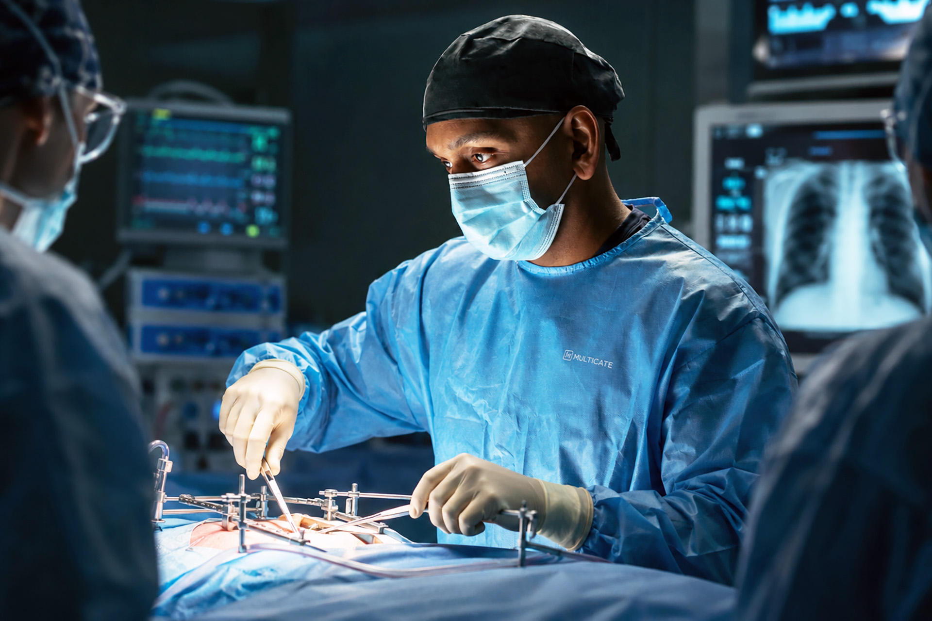
During a thoracotomy, the surgeon opens the chest through an incision between the ribs to perform the planned operation.
The operation is done under general anaesthesia.
You are usually positioned on your side so the surgeon can reach the affected part of the chest.
A breathing tube is placed to help control breathing during surgery.
In many thoracic operations, one lung is temporarily deflated to give the surgeon space to work safely.
The thoracotomy incision is usually made on the side or back of the chest, sometimes extending towards the anterior chest wall or following the posterior axillary line for an anterolateral thoracotomy.
In a posterolateral thoracotomy, the surgeon usually makes a cut between the ribs on the side or back of the chest to reach the lung or pleural space. The exact position depends on which part of the chest needs treatment and the type of operation planned.
Once the chest is open, a rib spreader may be used to gently separate the ribs and improve access. This gives the surgeon a clearer view of the chest cavity and exposes the mediastinal pleura, pulmonary ligament and pulmonary hilum when lung surgery is planned.
The thoracic aorta may also be exposed in selected operations. Because the rib edges can be sharp during incision and exposure, careful technique is needed to reduce the risk to the operating team and protect nearby nerves and blood vessels.
The surgeon then performs the planned treatment. This may include removing part of the lung, repairing injury, removing tumour, draining infection, taking tissue samples, removing thick pleural scar tissue, or controlling bleeding.
In some emergency trauma cases, the aorta may be cross-clamped, meaning the main blood vessel from the heart is temporarily compressed to reduce bleeding below the chest and redirect blood flow towards vital organs such as the heart and brain. This can be lifesaving, but it can also reduce blood flow to organs farther down the body and may cause ischaemia, which means tissue injury from reduced blood supply.
At the end of the operation, one or more chest drains are usually placed. These tubes remove air, blood or fluid from the chest while the lung re-expands.
The intercostal space is reapproximated, and the incision is then closed with stitches, staples or surgical glue.
The length of surgery varies. Some procedures take a few hours. More complex cancer, trauma or pleural operations may take longer.
What Happens after a Thoracotomy?
After a thoracotomy, you are monitored closely while your breathing, pain control, chest drains and lung expansions are assessed.
You may wake up in a recovery area, high-dependency unit, or intensive care unit. You may have oxygen, a drip, a urinary catheter, monitoring wires, and one or more chest drains.
Pain control is important after a thoracotomy because the operation involves the chest wall and ribs. Pain relief may include tablets, injections, nerve blocks, an epidural, or patient-controlled pain medicine. Good pain control helps you breathe deeply, cough, and walk, and reduces the risk of pneumonia.
Transcutaneous electrical nerve stimulation, also called TENS, may help manage post-thoracotomy pain in selected patients. Persistent pain after thoracotomy may need specialist review.
Chest drains often remain for at least one to two days. They are removed when air leakage has settled, fluid drainage has reduced, and the lung has expanded.
Chest X-rays are used to check lung expansion. Nurses and physiotherapists will encourage breathing exercises, coughing, sitting up in bed, and walking as soon as it is safe.
Hospital stays depend on the operation, lung expansion, pain control and the occurrence of complications. Many patients stay about one week after a thoracotomy, although this can be shorter or longer depending on the operation and the patient’s recovery.
Uniportal VATS uses a single small incision in selected cases.
Robotic-Assisted Thoracic Surgery (RATS) uses robotic instruments controlled by the surgeon from a console to perform selected chest operations through small incisions.
Other cardiothoracic surgery techniques may be used for specialised heart and chest procedures, depending on the organ involved and the complexity of the disease.
Medical thoracoscopy uses a camera inserted through a small chest incision to inspect the pleura, take biopsies, drain fluid, or perform talc pleurodesis in selected patients.
Image-guided biopsy uses CT or ultrasound scans to guide a needle into an abnormal area so a tissue sample can be taken without open surgery.
Drainage procedures use a needle, catheter or chest tube to remove air, fluid, blood or pus from the pleural space.
Non-surgical treatment may include medicines, monitoring, oxygen therapy, antibiotics, chemotherapy, immunotherapy, targeted therapy, or radiation therapy, depending on the condition being treated.
Benefits and Expected Outcomes of a Thoracotomy
The main benefit of a thoracotomy is that it gives direct access to the chest when major treatment or complex surgery is needed.
An open thoracotomy and chest thoracotomy procedures allow the surgeon to see and reach the affected area clearly. This can be important when removing lung cancer, managing severe infection, controlling bleeding, treating major trauma or dealing with thick pleural scarring.
Expected outcomes depend on the condition being treated:
In lung cancer, a thoracotomy may allow removal of cancer when surgery is appropriate.
In empyema or trapped lung, it may help clear an infection and allow the lung to re-expand.
In trauma, it may control bleeding or repair an injury.
In pleural disease, it may provide diagnosis, symptom relief or control of recurrent fluid or air.
A thoracotomy may not treat the entire disease process on its own. Some patients need further treatment, such as antibiotics, chemotherapy, immunotherapy, targeted therapy, radiation therapy, pulmonary rehabilitation or long-term follow-ups.
In an emergency surgery, the expected outcome depends heavily on the injury, how quickly treatment begins, and whether the heart, major vessels or lungs can be repaired.
Risks and Possible Complications of a Thoracotomy
A thoracotomy is major surgery and can cause complications such as pain, bleeding, infection, air leak, pneumonia and breathing problems.
Pain is common because the incision involves the chest wall and tissues between the ribs. Some patients develop longer-lasting nerve pain or numbness near the incision, known as post-thoracotomy pain syndrome.
Other risks include wound infections, chest infections, prolonged air leaks, fluid build-ups, blood clots, irregular heartbeats, respiratory failures, poor lung expansions, reactions to anaesthesia, and the need for further procedures.
Damage to the phrenic nerve can also occur. This nerve helps control the diaphragm, an important breathing muscle.
In lung surgery, removing the lung tissue can carry added risks, especially for patients with reduced lung functions.
In an emergency thoracotomy, aortic cross-clamping can reduce the blood flow to organs below the clamp and may cause organ injury.
Neurological complications may also occur after severe traumas, often because of the original injury and reduced blood flow.
Risks are higher in patients with severe lung disease, heart disease, infection, frailty, poor nutrition, smoking history, or advanced cancers. Your surgeon will explain your personal risk based on your scans, test results and overall health.
Recovery and Follow-Up of a Thoracotomy
Recovery after a thoracotomy usually takes weeks to months, depending on the operation and the patient’s overall health.
Hospital recovery focuses on pain control, chest drain care, breathing exercises, walking and preventing complications. You may feel tired, sore and short of breath with activity at first. This usually improves gradually.
At home, you may be advised to walk daily, continue breathing exercises, take pain medications as prescribed, keep the wound clean and dry, and avoid heavy lifting until cleared.
Driving, work and exercise should only resume when your surgeon says it is safe.
Many patients need to be off work for about two months, although this depends on the type of work, the surgery performed and the speed of recovery.
Follow-ups may include wound reviews, chest X-rays, lung function assessments, pathology results and treatment planning.
If any tissue was removed, the laboratory results guide the next steps. If a cancer is found, care may involve oncology as well as thoracic surgery.
Seek urgent medical review if you develop a fever, worsening breathlessness, increasing chest pain, wound redness, swelling, pus, heavy bleeding, dizziness, fainting, coughing up large amounts of blood, or sudden worsening after discharge.
How Neumark Can Help
Neumark Lung & Chest Surgery Centre can help determine whether a thoracotomy is needed or whether a minimally invasive option is more suitable.
Neumark specialises in minimally invasive thoracic surgery with a multidisciplinary approach led by Dr Harish Mithiran, senior consultant thoracic surgeon at Gleneagles and Mount Alvernia hospitals. For patients with lung tumours, pleural disease, empyema, trapped lung, pneumothorax, malignant pleural effusion or complex chest conditions, this means careful assessment before recommending an operation.
Contact Neumark today for a consultation or second opinion.
The purpose of a thoracotomy is to give the surgeon direct access to the chest cavity. It may be used to remove lung tissue, treat cancer, drain an infected area, repair an injury, obtain a biopsy, or manage complex pleural disease.
A thoracotomy is open chest surgery through an incision between the ribs. A thoracostomy usually refers to creating a small opening to place a chest tube to drain air, fluid, blood or pus.
Survival after a thoracotomy depends on the reason for surgery, the patient’s health, the type of operation and whether the surgery is planned or an emergency. A planned thoracotomy for selected patients carries different risks from an emergency thoracotomy after severe trauma.
A thoracotomy can be painful because it involves the chest wall and tissues between the ribs. Pain control is an important part of recovery and may include tablets, injections, nerve blocks or an epidural.
Yes. A thoracotomy refers to open surgery through the chest wall. VATS, U-VATS and RATS are minimally invasive alternatives that use smaller incisions in suitable patients.
Many patients need several weeks to recover, and full recovery may take months depending on the operation. Recovery is affected by pain control, lung function, age, general health and whether complications occur.
DISCLAIMER: The information provided on this website is for general informational purposes only and is not intended as a substitute for professional medical advice, diagnosis, or treatment. The use of this website does not create a doctor-patient relationship and no medical advice should be inferred or assumed. It is the user’s sole responsibility to seek the advice of their healthcare professionals for any medical concerns they may have and the user should not disregard, or delay, prompt medical advice for any such condition.
latest insights
Read our latest insights on lung diseases and chest conditions, including prevention tactics, treatments, and more.