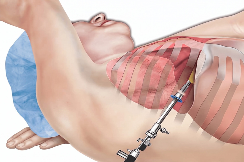
A thoracoscopy allows doctors to view the pleural space, drain fluid, take biopsies, and treat selected chest conditions through small incisions.
What Is a Thoracoscopy?
A thoracoscopy is a minimally invasive procedure that lets a thoracic surgeon look inside the chest using a thin tube with a camera and light called a thoracoscope.
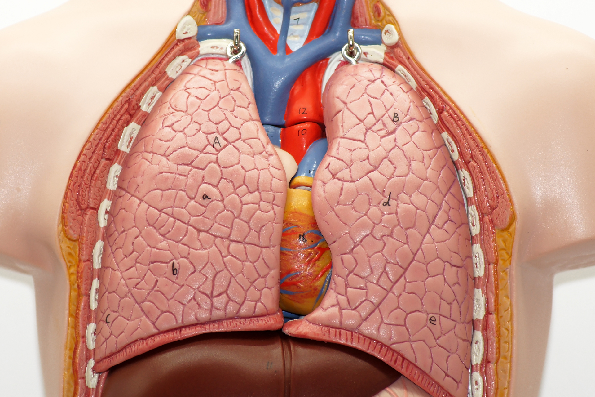
The camera is passed through a small incision in the chest wall to examine the pleura, the thin lining that covers the outside of the lung and the inside of the chest wall. The space between these layers is called the pleural space or pleural cavity, which contains a small amount of fluid.
A thoracoscopy may be diagnostic, therapeutic, or both:
Diagnostic thoracoscopy is used to identify the cause of symptoms, obtain pleural tissue samples, or evaluate abnormal imaging findings.
Therapeutic thoracoscopy is used to treat a problem, such as draining the pleural space, applying talc pleurodesis to reduce recurrence, or treating selected pleural disease.
Video-Assisted Thoracoscopic Surgery (VATS) is a surgical form of thoracoscopy performed by a thoracic surgeon, usually under general anaesthesia. You may also hear the term medical thoracoscopy. This is usually performed by a respiratory or pleural specialist using local anaesthesia and sedation.
Schedule a Consultation
Why Is a Thoracoscopy Performed?
A thoracoscopy is performed when doctors need to directly visualise the pleural space, obtain tissue samples, drain the space, or treat recurrent collections of pleural fluid around the lung.
It is commonly used when pleural collections continue to accumulate, and the cause remains unclear after scans and tests. This is sometimes described as a thoracoscopy pleural effusion assessment. It is especially useful when cancer, tuberculosis, chronic pleural infection, mesothelioma, or another pleural disease is suspected.
Pleural tissue sampling when fluid tests are inconclusive
Talc pleurodesis to reduce recurrence
In selected cases, VATS can also be used to remove tumours, excise damaged lung tissue, remove blebs, and treat conditions such as emphysema.
In cancer care, a thoracoscopy may help confirm the diagnosis, support staging, relieve symptoms, or guide treatment such as chemotherapy, immunotherapy, targeted therapy, or radiation therapy. It does not cure most advanced cancers on its own, but can provide important information and improve breathing comfort.
When to See a Thoracic Surgeon
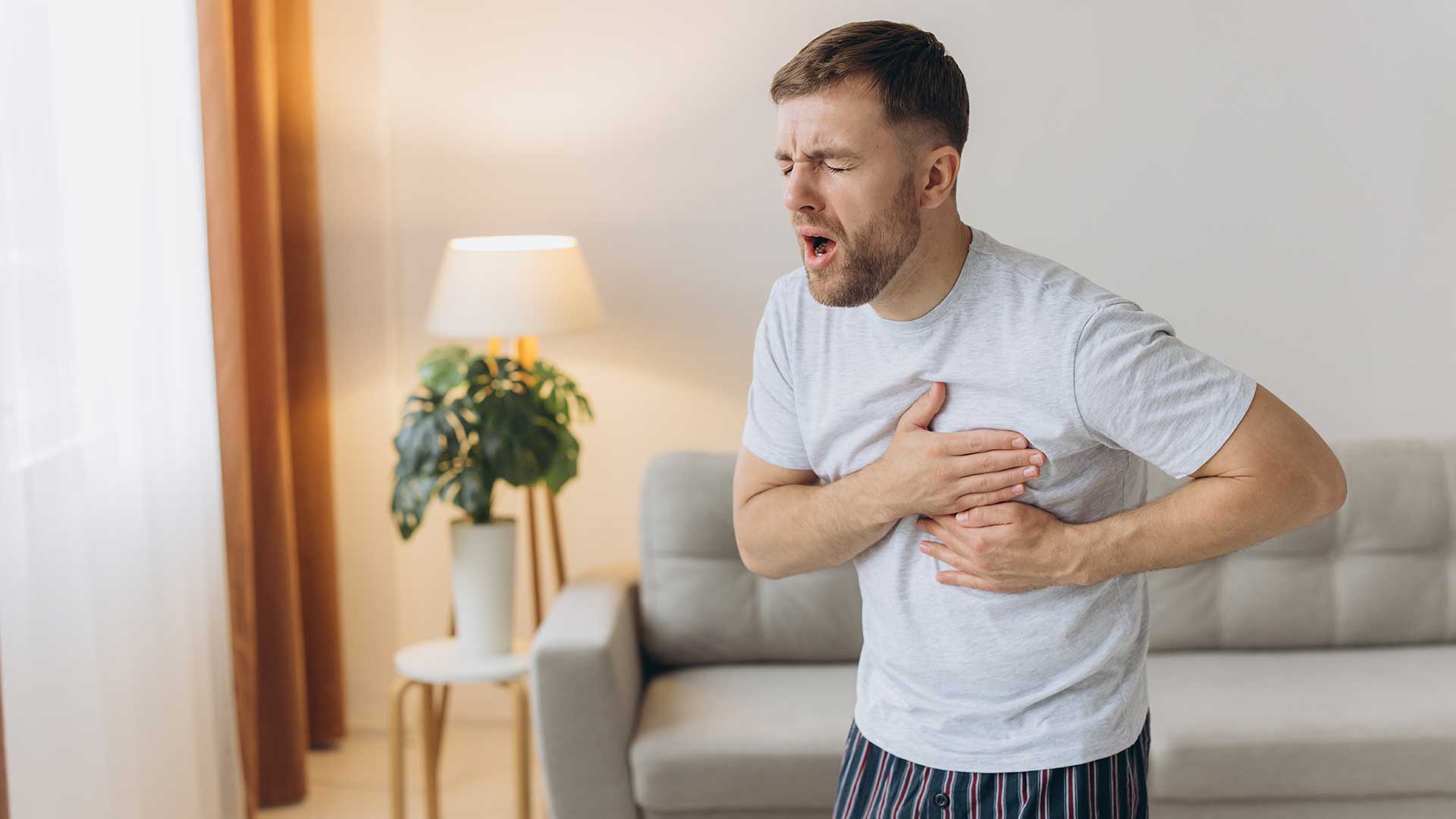
You should see a thoracic surgeon when pleural fluid, pleural thickening, recurrent pneumothorax or unexplained chest symptoms need direct assessment.
A lung specialist review is important if you have breathlessness, chest pains, recurrent pleural effusions, brownish pleural fluids, an unexplained weight loss, fever, a persistent cough, or imaging showing pleural nodules or thickening. It is also important if the previous thoracentesis did not provide a clear diagnosis.
A thoracic surgeon may be needed when VATS is indicated. This procedure may include:
More complex tissue sampling
Treatment of trapped lung
Management of empyema
Surgical pleurodesis
Recurrent pneumothorax surgery
Decortication
Deciding between thoracoscopy and VATS depends on which approach is best suited to the patient’s diagnosis, anatomy, fitness, and treatment goals. A medical thoracoscopy may be sufficient for a pleural inspection, biopsy, and talc pleurodesis in selected patients. VATS with a video monitor may be better when more extensive surgical treatment is needed.
Schedule a Consultation
How to Prepare for a Thoracoscopy
Preparation for a thoracoscopy focuses on confirming the reason for the procedure and reducing the risks of bleeding, infection and anaesthesia.
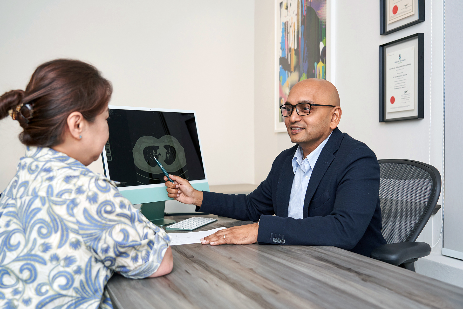
Before the procedure, your doctor will review your symptoms, scans, medical history, medications and allergies.
You may need blood tests, a chest X-ray, an ultrasound, a CT scan, lung function tests, a heart assessment, or pleural sampling tests.
You may be asked not to eat or drink for several hours before the thoracoscopy. A few hours of fasting helps reduce anaesthesia complications.
Blood-thinning medicines are often stopped at least 5 days before the procedure, on medical advice. Stop taking your blood-thinning medication as directed and other regular medicines only if your team advises you to.
You should also arrange for someone to drive you home if a same-day discharge is planned.
If a medical thoracoscopy is planned, you may receive sedation and a local anaesthetic.
If a VATS is planned, you will usually have a general anaesthetic, which means you are asleep during the operation.
Bring previous scan reports, test results and a medication list to your consultation. These will help your doctor decide whether a thoracoscopy is likely to give useful information, or whether another test would be safer.
What Happens During a Thoracoscopy?
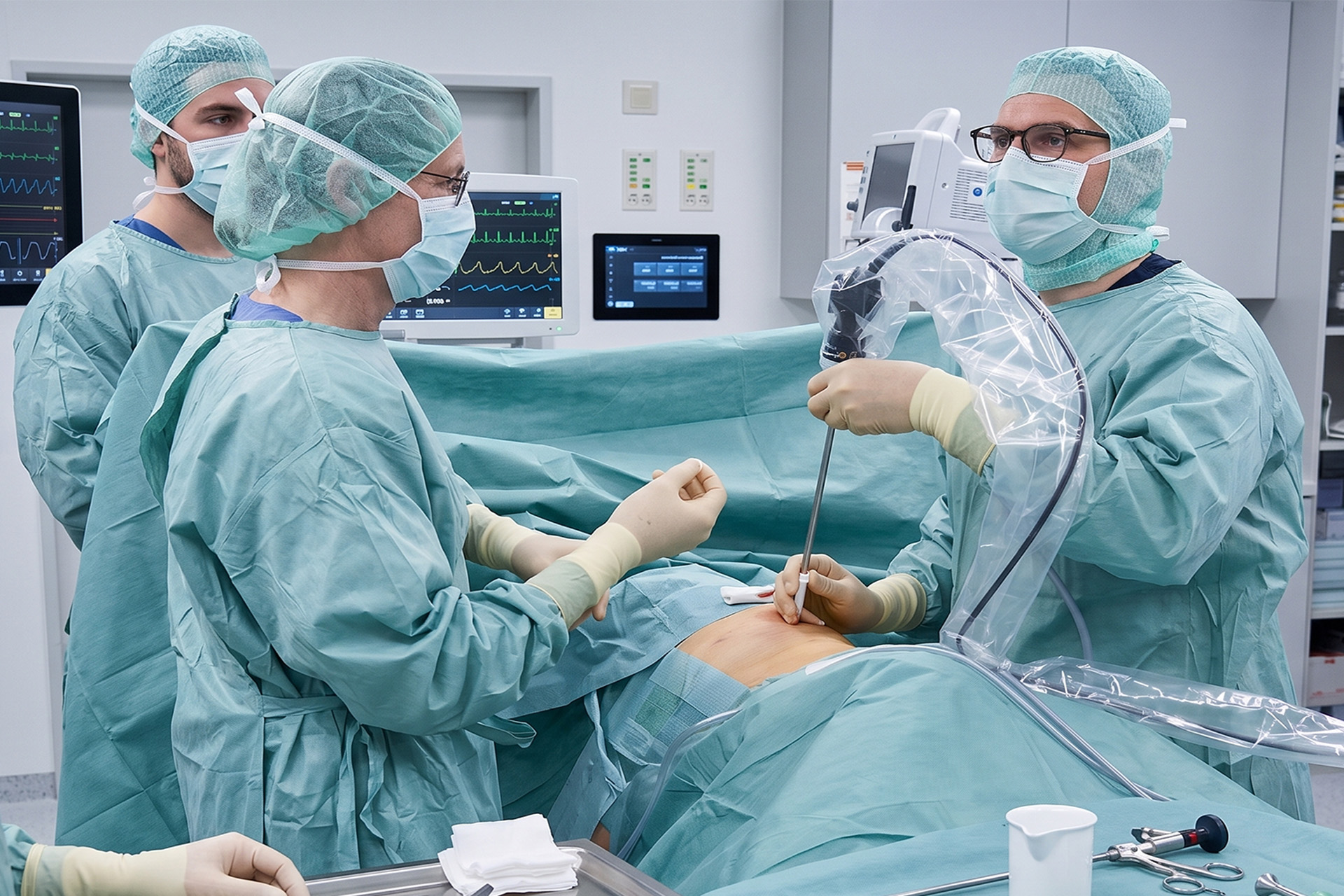
During a thoracoscopy, a camera is placed through a small chest incision so the doctor can inspect the pleura, drain fluid and take biopsies if needed.
You are usually positioned on your side.
Oxygen levels, blood pressure, pulse and breathing are monitored throughout.
Local anaesthesia is used to numb the chest wall for a medical thoracoscopy. Sedation may be given to help you relax. There is a small risk of allergic reaction, and other possible risks are explained beforehand. For VATS, general anaesthesia is used in the operating room.
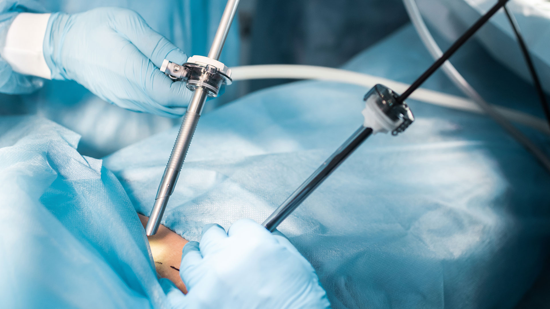
The doctor makes one or more small cuts in the chest wall, each of which is closed with a small stitch afterwards.
The thoracoscope is inserted through the opening.
If pleural fluid is present, it can be drained.
The pleura is inspected carefully. Areas that look abnormal can be biopsied, meaning small tissue samples are removed for laboratory testing.
Samples of lung tissue or lymph nodes may also be taken. Taking samples helps confirm whether cancer cells are present and may identify lung cancer.
If talc pleurodesis is needed, sterile talc may be applied to the pleural space to help the lung lining adhere to the chest wall. This can reduce the chance of recurrence.
If an indwelling pleural catheter is needed, it may be placed during the same procedure in selected patients.
A thoracoscopy may take about 40 to 60 minutes for simpler medical procedure cases. Video-Assisted Thoracoscopy may take longer if surgery, decortication or treatment of leaks is needed. Both are safe procedures.
What Happens after a Thoracoscopy?
After a thoracoscopy, you are monitored while the sedation or anaesthesia wears off and while the lung and pleural space are checked.
You may feel sleepy, tired or mildly sore.
If a breathing tube was used during general anaesthesia, you may have a sore throat or hoarse voice for a short time.
Chest discomfort is common, especially if talc pleurodesis, a chest tube or chest drain is used.
A chest drainage bottle may be left in place to remove air or blood from the pleural space. The chest drain bottle often stays in for 1 to 2 days after the procedure, but it may be needed longer if drainage continues or there is an air leak.
A chest X-ray is usually done to check that the lung has re-expanded and that the drain is in the correct position.
Some go home the same day after a medical thoracoscopy; others stay overnight in the hospital. Most patients who have undergone a surgical thoracoscopy are discharged from the hospital within 2 to 4 days, depending on the procedure, chest cavity drain, lung re-expansion status, hospital review and recovery.
Hospital nursing staff monitor for a few days, with rechecks over the same period as drains are removed.
The hospital team gives specific instructions and further information for at-home care.
Benefits and Expected Outcomes of a Thoracoscopy
The main benefit of a thoracoscopy is that it can provide diagnosis and treatment through small incisions.
A thoracoscopy allows direct visual inspection of the pleura. This can make tissue sampling more accurate than blind methods. It can also drain pleural collections and excess fluid, support pleurodesis, assess lung entrapment, and help guide further treatment.
Compared with traditional open chest surgery, a thoracoscopy usually causes less postoperative pain, only a small amount of bruising, and allows a quicker recovery. Cosmetic results improve because the scars are smaller. The unaffected side of the chest remains undisturbed.
For patients with pleural fluid, the expected benefits may include a clearer diagnosis, improved breathing, and fewer repeat drainage procedures if pleurodesis is successful.
For patients with suspected lung cancer, a thoracoscopy may confirm the cancer type and support treatment planning.
For a pneumothorax, VATS may help reduce recurrence by treating blebs and performing pleurodesis or a pleurectomy where appropriate.
The result depends on the underlying condition. A thoracoscopy can provide answers and symptom relief, but it may not remove the need for oncology treatment, antibiotics, long-term catheter care or follow-up imaging.
Risks and Possible Complications of a Thoracoscopy
A thoracoscopy is a generally safe procedure, but possible complications include pain, bleeding, infection, air leak, pneumothorax and fluid recurrence.
Pain is common after the procedure, especially around the incision or chest drain site.
Bleeding can occur after sampling; a little blood in the chest is sometimes seen.
Infection may affect the wound, pleural space or lung.
Air may leak into the pleural space or thoracic cavity and cause pneumothorax. It may also return if the underlying disease remains active.
Other possible risks include fevers, low oxygen levels, reactions to sedation or anaesthesia, prolonged air leaks, chest drain discomforts, pneumonia, blood clots and the need for further treatment.
Our surgeons and care team at Neumark Lung & Chest Surgery Centre will explain the side effects based on your diagnosis, age, lung function, other medical conditions and the planned medical procedure.
Recovery and Follow-Up after a Thoracoscopy
Recovery after a thoracoscopy usually takes days to weeks, depending on the type of procedure and the condition being treated.
Many patients can start normal activities within 2 weeks of the procedure, while full recovery may take 4 to 6 weeks, especially after a VATS.
Most patients return to light activities shortly after a medical thoracoscopy.
You may be advised to walk regularly, do breathing exercises, take pain medicine and any prescribed medicine as instructed, and keep the wound clean and dry.
Avoid heavy lifting and strenuous activity until your doctor says it is safe. Your doctor will advise on when to resume.
If you go home with an indwelling pleural catheter, you will be taught how drainage works and when to call your doctor for help, if needed
Follow-up after the procedure may include biopsy results, a wound check, a chest X-ray, and a discussion of further treatment.
Tissue results may take several days after the procedure. If cancer is found, your care may involve oncology, respiratory medicine and thoracic surgery.
Seek urgent medical review if you develop fever, worsening breathlessness, increasing chest pain, wound redness, swelling, pus, heavy bleeding, dizziness, blue lips, or sudden deterioration.
How Neumark Can Help
Neumark Lung & Chest Surgery Centre can help determine whether a thoracoscopy, medical thoracoscopy, VATS or another pleural procedure is the right next step.
Neumark specialises in minimally invasive thoracic surgery with a multidisciplinary approach led by Dr Harish Mithiran, senior consultant at Gleneagles and Mount Alvernia hospitals. For patients with pleural conditions, pneumothorax, pleural thickening, suspected mesothelioma, empyema or trapped lung, this means careful reviews before recommending a procedure.
If a thoracoscopy has been recommended or if you have an unexplained effusion around the lung, contact Neumark for a consultation.
A thoracoscopy is minimally invasive, but it is still a chest procedure. A medical thoracoscopy is usually less invasive than VATS. VATS is a surgical form of thoracoscopy and is usually done under general anaesthesia.
You should not feel pain during the procedure because local anaesthetic, sedation or general anaesthesia is used. Chest discomfort can occur afterwards, especially if a chest drain or talc pleurodesis is used. Pain relief is provided.
A thoracoscopy uses small cuts and a camera to look inside the chest. A thoracotomy is open chest surgery through a larger incision. A thoracotomy may be needed when the disease is too complex to treat safely through small incisions.
A simpler medical thoracoscopy may take about 40 to 60 minutes. VATS can take longer if a biopsy, pleurodesis, decortications, or other surgical treatments are needed.
Common side effects include chest soreness, tiredness, a mild fever after talc pleurodesis, and discomfort from a chest drain. Less common complications include bleeding, infection, pneumothorax, air leak or fluid recurrence.
A thoracoscopy can help prevent recurrent pleural fluid when talc pleurodesis is performed, and the lung can fully expand. It may not work as well if the lung is trapped or the underlying disease remains very active.
DISCLAIMER: The information provided on this website is for general informational purposes only and is not intended as a substitute for professional medical advice, diagnosis, or treatment. The use of this website does not create a doctor-patient relationship and no medical advice should be inferred or assumed. It is the user’s sole responsibility to seek the advice of their healthcare professionals for any medical concerns they may have and the user should not disregard, or delay, prompt medical advice for any such condition.
latest insights
Read our latest insights on lung diseases and chest conditions, including prevention tactics, treatments, and more.