

Our Services

Pleurodesis

Pleurodesis seals the space around the lung to help prevent repeated fluid build-up or lung collapse. Learn what to expect.

What Is Pleurodesis?

Pleurodesis is a procedure that produces controlled adhesion between the pleural layers, sealing the pleural space around the lung to help prevent fluid or air from collecting there.

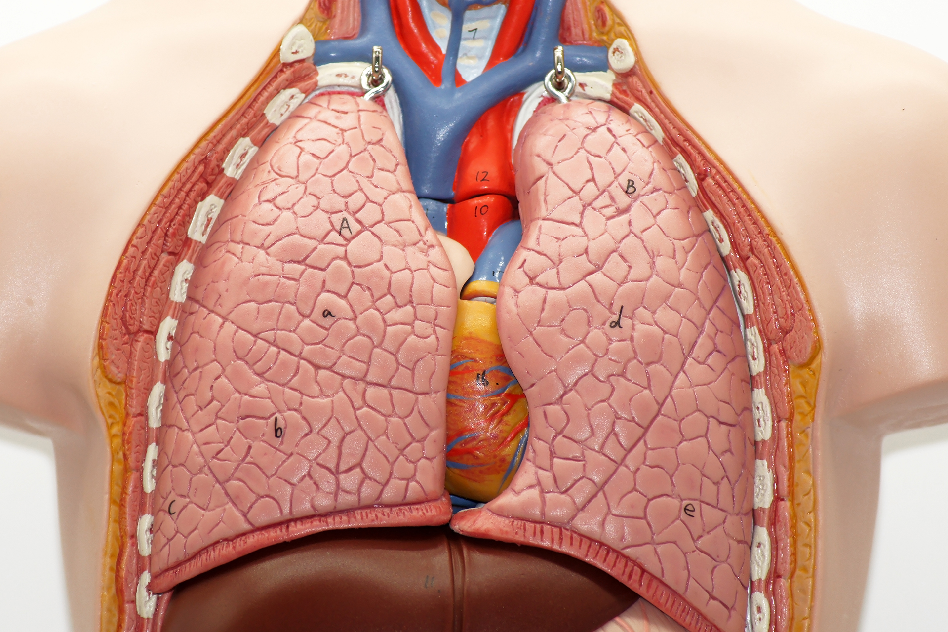
The lungs are covered by a thin lining called the visceral pleura. The inside of the chest wall is lined by the parietal pleura. The small gap between these layers is called the pleural space. Pleural space anatomy along the chest wall determines how fluid or air collects within it. Normally, this space contains only a small amount of fluid to help the lung move smoothly during breathing.

Pleurodesis works by causing the two pleural layers to adhere. Once the space is sealed, there is less room for fluid or air to return. This can help patients with recurrent pleural effusion, repeated pneumothorax, or selected cancer-related fluid around the lung.

Pleurodesis does not treat every underlying disease. For example, in malignant pleural effusion, it does not cure the cancer. Its main aim is to control the symptoms, reduce recurrence, and help keep the lung expanded. In pneumothorax, the aim is to lower the risk of another lung collapse.

There are different types of pleurodesis.

  • Chemical pleurodesis uses a medicine or irritant, most commonly sterile talc, to induce controlled inflammation, causing the pleural layers to seal across the pleural surfaces.
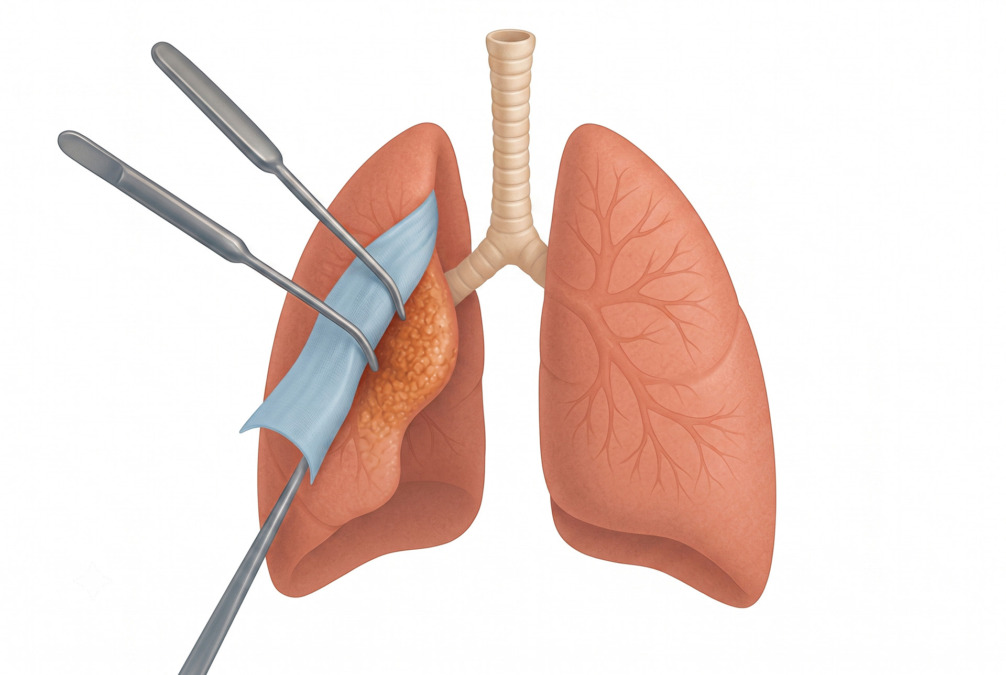
  • Mechanical pleurodesis uses mechanical abrasion to roughen the pleural surface so the pleura heals together. Mechanical methods include scuffing and gauze abrasion, which aim to promote adhesion between the pleural layers.

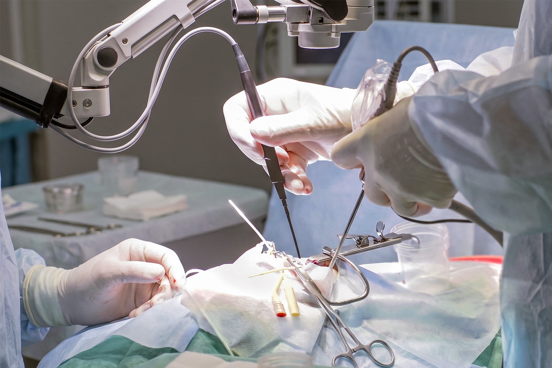
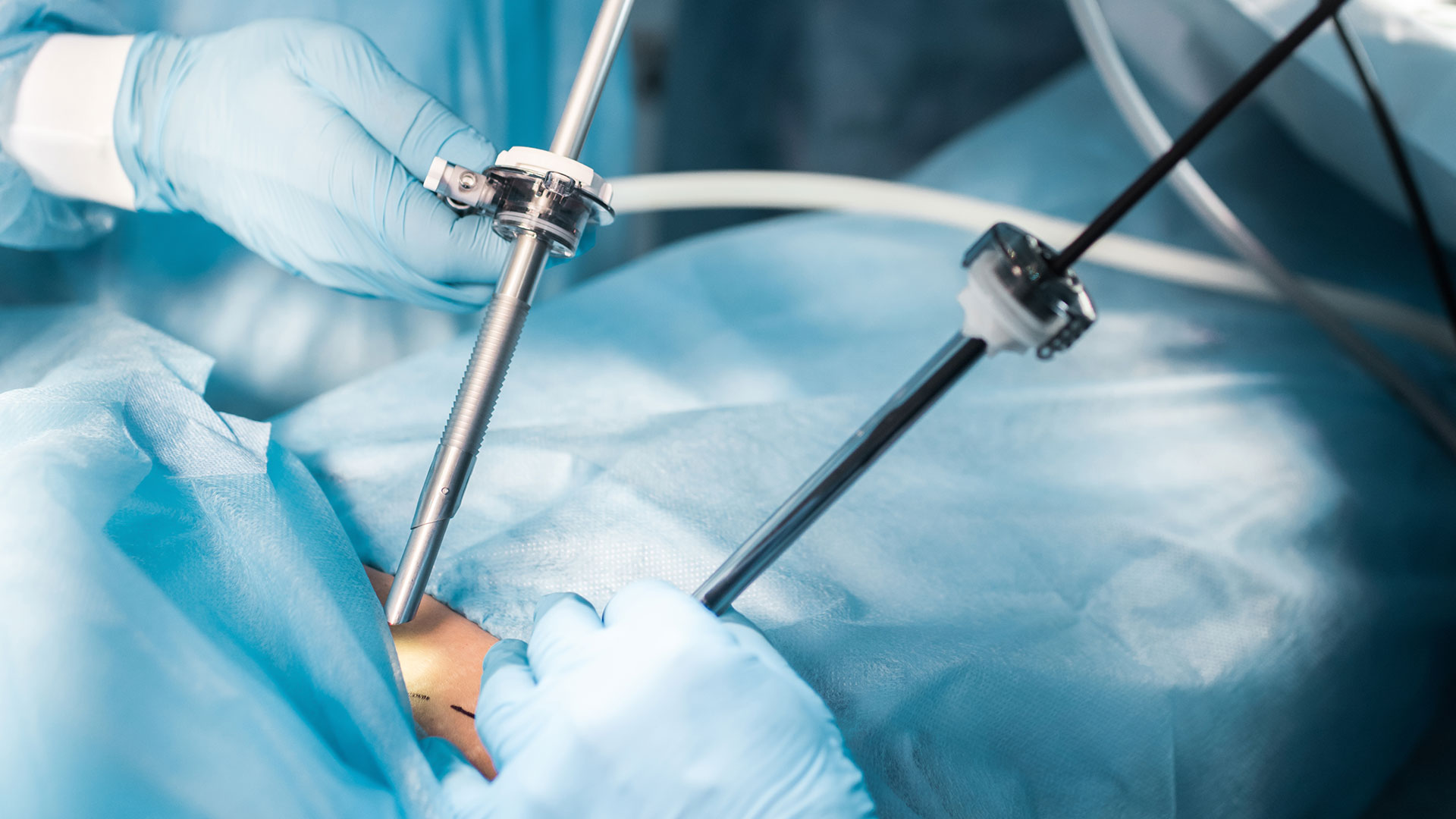
Surgical pleurodesis may be done through Video-Assisted Thoracoscopic Surgery (VATS) or through open thoracotomy in selected cases.

pleurodesis

Primary Conditions Treated with Pleurodesis

Pleurodesis is mainly used to treat recurrent pleural effusions and recurrent pneumothoraces.

  • Pleural Effusion: A pleural effusion is fluid around the lung. It may be caused by cancer, pleural infections, heart failure, liver disease, kidney disease, inflammatory disease, or other conditions. Heart failure is among the most common causes of bilateral pleural effusion. Pleurodesis may be considered when the fluid keeps returning and causing breathlessness.
  • Malignant Pleural Effusion: A malignant pleural effusion is fluid around the lung caused by cancer. The fluid may press on the lung, making breathing difficult. In selected patients, pleurodesis for malignant pleural effusion can reduce the recurrent fluid accumulation and the need for repeated drainage procedures. For pleurodesis to work well, the lung usually needs to re-expand after the fluid is drained.
  • Pneumothorax: A pneumothorax is air around the lung that causes part or all of the lung to collapse. Spontaneous pneumothorax can occur without trauma, and primary spontaneous pneumothorax often affects tall, young people. Secondary spontaneous pneumothorax occurs in those with lung disease. Pleurodesis may be recommended for recurrent pneumothorax, primary spontaneous pneumothorax with recurrence, persistent air leaks, or patients at higher risk.
  • Cancer-Related: Cancer can cause fluid to build up around the lung. This is called a malignant pleural effusion and may happen with lung cancer, breast cancer, lymphoma, ovarian cancer, mesothelioma, or other cancers affecting the pleura. Pleurodesis may be considered when the fluid keeps returning after drainage.

Pleurodesis indication for individual patients depends on the cause, symptoms, lung expansion in the pleural space, expected benefit and overall health. It is not suitable for all patients with pleural fluid or a pneumothorax. If the lung is trapped and cannot expand, pleurodesis is less likely to succeed because the pleural layers cannot come into contact properly.
mechanical pleurodesis


When to See a Thoracic Surgeon

You should see a thoracic specialist if fluid or air around the lung keeps returning, does not drain well, or causes persistent breathlessness.

A specialist review and thoracic surgery assessment are important when pleural effusion or pneumothorax has occurred more than once. It is also important if a chest drain has not solved the problem, if the lung does not re-expand, or if scans suggest trapped lung, pleural thickening, infection, cancer or loculated fluids.

Neumark Lung & Chest Surgery Centre’s specialists can assess whether pleurodesis surgery is appropriate and whether it should be performed via chest tube, medical thoracoscopy, VATS pleurodesis, uniportal VATS, or another approach. The best option depends on the diagnosis, the patient’s anatomy, disease stage, lung expansion, treatment goals, and the patient’s fitness for anaesthesia.

In Singapore, timely thoracic surgery assessment can help patients avoid repeated emergency drainage, prolonged symptoms, and delays in cancer or pleural disease treatment.

surgical pleurodesis

How to Prepare for Pleurodesis

Preparation for pleurodesis focuses on confirming the cause, checking lung expansion, and making the procedure as safe as possible.

  • Before pleurodesis, your doctor will review your symptoms, scans, fluid test results, medical history, and current medicines.
  • Blood thinners may need to be adjusted, but only with medical advice.
  • You may need blood tests, chest X-rays, an ultrasound, CT scans, lung function tests, or a heart assessment, depending on your condition.

If fluid is present, a sample may be tested for infection, cancer cells, protein, glucose, pH, cell count, and other markers. Pleurodesis efficacy depends on pleural fluid biochemistry, and such tests guide the patient’s treatment.

If the diagnosis is unclear, a pleural biopsy may be needed before or during the procedure.

The lung must usually be able to expand after fluid or air is drained. If the lung remains trapped, pleurodesis may not work well. In that case, alternatives such as an indwelling pleural catheter or decortication may be considered.

You may be asked not to eat or drink for several hours before surgical pleurodesis or VATS pleurodesis. Your care team will explain what medicines to take, what to stop, and whether you should expect local anaesthesia, sedation, or general anaesthesia. Your doctor may also advise avoiding non-steroidal anti-inflammatory drugs (NSAIDs) after pleurodesis because these medicines reduce inflammation, which may affect how well the pleural layers seal.


What Happens During Pleurodesis?

During the procedure, the pleural space is drained and then treated so that the lung lining adheres to the chest wall.

  1. Anaesthesia or sedation is given. Chemical pleurodesis through a chest tube may use local anaesthesia and pain control, with or without light sedation, to manage the pain during the procedure. VATS pleurodesis is usually done in the operating room under general anaesthesia.
  2. Fluid or air is drained. Chest tube insertion is performed if one is not already in place. Smaller chest tubes are often preferred for pleurodesis procedures as they can be effective with less pain than larger tubes. Chest tube size selection depends on the indication, and chest tube administration of the sclerosing agent follows once drainage is adequate.
  3. The doctor checks the lung expansion. Pleurodesis works best when the lung expands sufficiently for the pleural layers to come into contact.
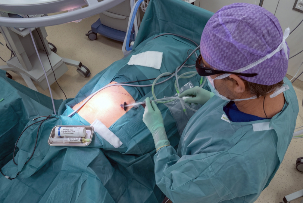
  4. The pleural space is treated. In chemical pleurodesis, sterile talc is the most common agent. Talc slurry may be given through a chest tube. Talc poudrage is performed during a thoracoscopy by spraying talc powder. Other agents have been used in selected situations, including doxycycline, bleomycin pleurodesis, silver nitrate, povidone-iodine, and iodopovidone. In mechanical pleurodesis, the surgeon gently irritates the pleural surface during surgery.
  5. The medicine is distributed. If the talc slurry, mixed with sterile saline before use, is delivered through a chest tube, the tube may be clamped to allow the slurry to coat the pleural space. Talc slurry distribution depends on patient positioning. The chest drain is often reopened after about 3 hours, depending on the protocol used and the patient’s condition.
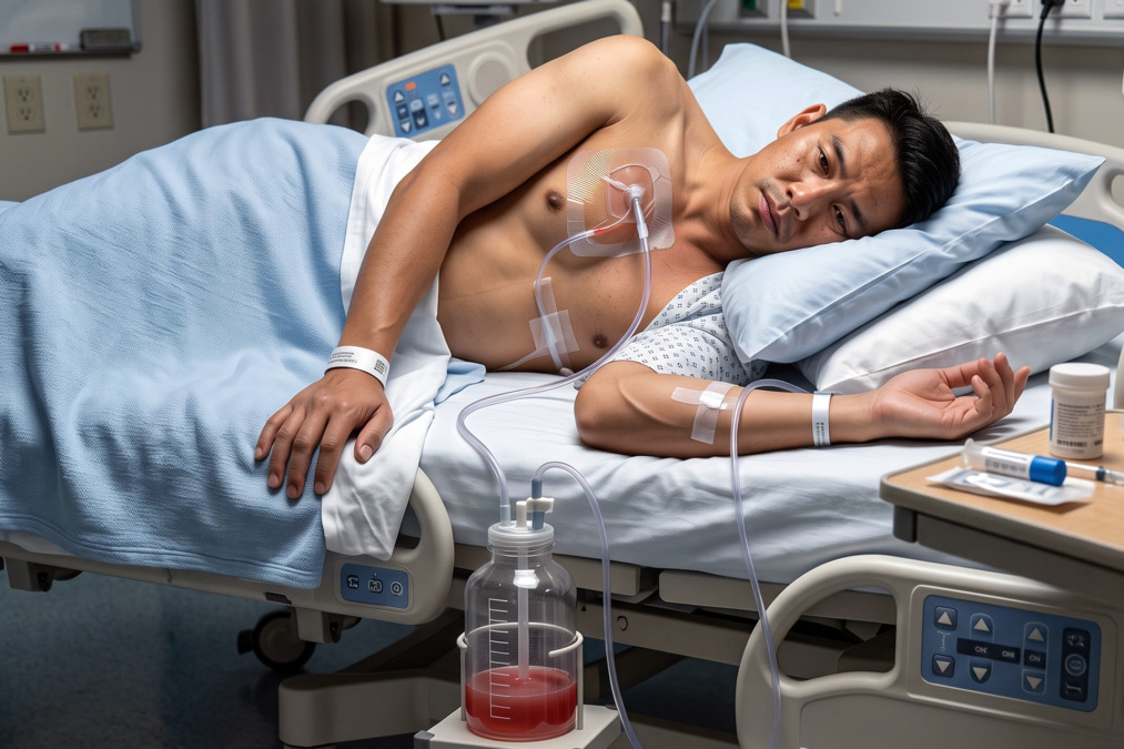
  6. A chest drain remains in place. The drain removes remaining fluid or air while the pleural layers begin to seal.

The pleurodesis procedure may take 30 minutes to a few hours in the operating room, depending on the method used and whether other procedures are done at the same time. Talc pleurodesis is a commonly performed medical procedure in this setting. 

If VATS is used, the surgeon may also inspect the pleura, take biopsies, treat air leaks, remove abnormal tissue, or perform partial pleurectomy if needed.


pleurodesis procedure


What Happens After Pleurodesis?

After pleurodesis, you are monitored while the lung stays expanded, and the chest drain removes the remaining fluid or air.

  • You may feel chest pain or tightness, a cough, fever, or tiredness for a short time. Pain relief is important because it helps you breathe deeply, cough and walk.
  • A chest X-ray is usually performed about 24 hours after the pleurodesis to confirm that the lung has expanded and that the chest tube is in the correct position. If the X-ray is satisfactory and drainage has reduced, the chest tube may be removed.
  • Some patients stay in the hospital for a few days. Others may need longer depending on the cause, the amount of drainage, infection risks, air leaks, cancer treatment needs, or the patient’s overall health.
  • If pleurodesis is done for malignant pleural effusion, your cancer care may continue after recovery. This may include chemotherapy, immunotherapy, targeted therapy, radiotherapy, or palliative care support, depending on the cancer type and treatment plan.
  • A follow-up chest X-ray is often recommended after 4 to 6 weeks. This helps confirm that the lung remains expanded and that fluid or air has not returned.

Risks and Possible Complications of Pleurodesis

Pleurodesis complications can include pain, fever, infection, bleeding, air leaks, failed pleurodesis, and breathing problems.

Pain is common among patients because pleurodesis induces controlled inflammation. Fever can also occur in some patients after chemical pleurodesis. The effects of this medical procedure are usually managed with medicines and close monitoring. Adverse events are uncommon overall.

Possible complications include wound infections at the chest wall, chest infections, bleeding, pneumothorax, persistent air leaks, low oxygen levels, fluid re-accumulation, or failure of the pleural layers to seal. Infection can occur if an aseptic technique is not maintained during the tube placement, medication administration, or drain care. Rare but serious complications of the procedure can include severe breathing difficulty or infection in the pleural space.

Acute respiratory distress syndrome (ARDS) has been reported in rare patients with talc use. This is uncommon, but it is one reason sterile, graded talc and careful patient selection are important.

Pleurodesis may not work if the lung cannot fully expand, if the fluid is loculated into separate pockets, if the pleural fluid pH or glucose is low, or if the underlying disease remains very active.

The risks are different for each individual patient. A frail patient with advanced cancer may face different risks from a young person having pleurodesis for pneumothorax. Your treating team will explain the likely benefits and risks based on your diagnosis and scans.


Recovery from Pleurodesis

Pleurodesis recovery usually takes days to weeks, depending on the method used and the condition being treated.

Recovery after a chemical pleurodesis through a chest tube may be shorter than recovery after a surgical approach. Recovery after VATS is often faster than open surgery, but this depends on the complexity of the disease and the patient’s general health.

During recovery, you may be advised to walk regularly, do breathing exercises, take pain medicine as prescribed, keep the wounds clean and dry, and avoid heavy lifting until cleared by your medical team. If you had VATS or another surgical approach, you may have small chest wounds that need simple dressing care.

Your team may advise avoiding NSAIDs and certain medicines unless they specifically say these are safe for you. Do not stop prescribed medicines without medical advice.

Follow-up may include a chest X-ray, a review of symptoms, a wound check, and a discussion of further treatment. If pleurodesis was done for cancer-related fluid, your care may also involve an oncologist. If it was done for a pneumothorax, follow-up focuses on lung expansions, air leak resolutions, and recurrence risks.

Seek urgent medical review if you develop a fever, worsening breathlessness, increasing chest pains, wound redness, swellings, pus, heavy bleeding, dizziness, blue lips, or a sudden worsening after your hospital discharge.


pleurodesis surgery


Alternatives to Pleurodesis

Treatment options as alternatives to pleurodesis include observation, repeated drainage, chest tube drainage, an indwelling pleural catheter, surgery for trapped lung, or the treatment of the underlying condition. These treatment options aim to remove excess fluid and protect respiratory function.

  • Observation may be enough if the symptoms are mild and the fluid or air is not worsening. Repeated thoracentesis can relieve breathlessness from pleural effusion and remove excess fluid, but it may become inconvenient and carries the risks of repeated procedures.
  • An indwelling pleural catheter is a soft tube that stays in the chest and allows fluid to be drained at home. It may be suitable for malignant pleural effusion, especially if the lung is trapped or the patient wants to avoid repeated hospital procedures.
  • Decortication may be considered when a thick pleural peel traps the lung and prevents expansion. This is different from pleurodesis. Decortication removes restrictive scar tissue. Pleurodesis seals the pleural space. By sealing the pleural space, recurrence is reduced.
  • For pneumothorax, options may include observation, supplemental oxygen, needle aspiration, chest tube drainage, medical pleurodesis via chest tube, or surgery to treat blebs and reduce recurrence. Pleurodesis may be combined with these treatments in selected patients.


How Neumark Can Help

Neumark Lung & Chest Surgery Centre helps patients decide whether the pleurodesis procedure is the right treatment for them and which approach is safest for their condition.

Neumark specialises in minimally invasive thoracic surgery with a multidisciplinary approach led by Dr Harish Mithiran, senior consultant thoracic surgeon at Gleneagles and Mount Alvernia hospitals. For patients with recurrent pleural effusions, recurrent pneumothorax, malignant pleural effusions, trapped lung, or complex pleural disease, careful assessment before treatment is recommended.

If you have repeated fluid or air around the lung, or if pleurodesis has been recommended, contact Neumark for a consultation.
FAQs about Pleurodesis

Pleurodesis can be done through a chest tube or as part of a surgery. Chemical pleurodesis through a chest tube is less invasive than surgical pleurodesis. VATS pleurodesis is still a surgical procedure and is usually done under general anaesthesia.

If pleurodesis is successful, the pleural space stays sealed long term. However, fluid or air can return if the pleurodesis does not fully work or if the underlying disease remains active.

Sterile talc is the most commonly used agent for chemical pleurodesis. Other agents, such as doxycycline or bleomycin, may be used in selected cases depending on local practice and the patient’s condition.

The main downsides include pain, fever, need for a chest drain, hospital stay, possible complications, and the chance that it may not work. It may also be unsuitable if the lung cannot expand fully.

Pleurodesis itself is not intended to reduce life expectancy. In cancer-related effusions, the overall outlook depends mainly on the type and stage of the cancer, not the pleurodesis procedure.

Pleurodesis can cause chest pain because it creates controlled inflammation. Pain is usually managed with medicine. Surgical pleurodesis may cause more discomfort than chemical pleurodesis via chest tube, especially in the first few days after the procedure.

DISCLAIMER: The information provided on this website is for general informational purposes only and is not intended as a substitute for professional medical advice, diagnosis, or treatment. The use of this website does not create a doctor-patient relationship and no medical advice should be inferred or assumed. It is the user’s sole responsibility to seek the advice of their healthcare professionals for any medical concerns they may have and the user should not disregard, or delay, prompt medical advice for any such condition.

latest insights

Read our latest insights on lung diseases and chest conditions, including prevention tactics, treatments, and more.